
Muscle · Lower limb
Adductor magnus: origin, insertion, action and innervation
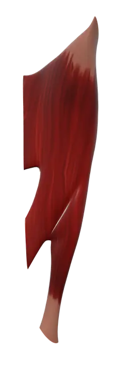
The adductor magnus is a thigh muscle from the ischiopubic ramus and ischial tuberosity to the femur. It adducts the thigh (obturator and tibial nerves).
Quick answer
- Latin name
- Musculus adductor magnus
- Origin
- Adductor part: inferior pubic ramus and ischial ramus. Hamstring part: ischial tuberosity (hip bone)
- Insertion
- Adductor part: gluteal tuberosity, linea aspera and medial supracondylar line. Hamstring part: adductor tubercle of the femur
- Action
- Adducts the thigh; the adductor part helps flex it, the hamstring part extends it
- Innervation
- Adductor part: obturator nerve, posterior division (L2–L4). Hamstring part: tibial division of the sciatic nerve (L4)
- Blood supply
- Deep femoral artery and its perforating branches, obturator artery, and the femoral artery
See it on the 3D body
The adductor magnus lit on the 3D hip and thigh, as in the hip and thigh quiz. Turn the body, peel the layers, tap any structure for its name.
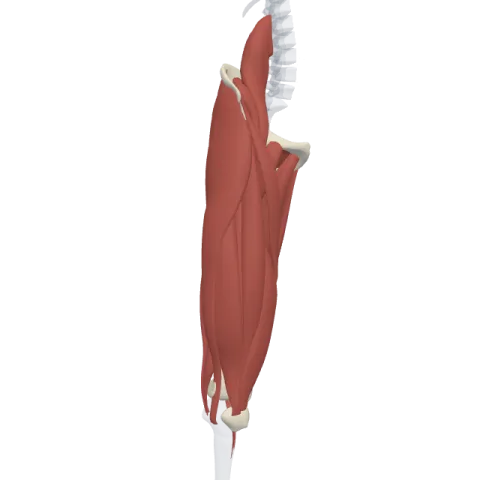
The adductor magnus is the largest and deepest muscle of the inner thigh, running from the ischiopubic ramus and ischial tuberosity to almost the whole length of the back of the femur. It adducts the thigh and has two parts with two nerves: the obturator nerve for the adductor part and the tibial division of the sciatic nerve for the hamstring part.
Where it is
The adductor magnus belongs to the medial compartment of the thigh. It is the deepest of the adductors and lies behind the adductor longus and the adductor brevis, forming a broad, thick partition between the adductor group and the hamstrings behind it.
Seen from behind, it fills the space between the medial hamstrings and the femur. It is large enough to be seen on the back of the thigh once the gluteus maximus and the hamstrings are removed. See how it fits with the other thigh muscles on muscles of the thigh.
Origin and insertion
The adductor magnus is really two muscles fused together, with different origins, insertions and nerves.
Adductor part (also called the pubofemoral part):
- Origin: the inferior pubic ramus and the ischial ramus of the hip bone.
- Insertion: a long line on the back of the femur, from the gluteal tuberosity down the linea aspera to the medial supracondylar line. Its upper, horizontal fibers are sometimes described separately as the adductor minimus.
Hamstring part (also called the ischiocondylar part):
- Origin: the ischial tuberosity, next to the hamstrings.
- Insertion: a strong, round tendon on the adductor tubercle, a small bump on top of the medial condyle of the femur, just above the knee.
Between the end of the adductor insertion and the tendon to the adductor tubercle there is a gap, the adductor hiatus. The femoral artery and vein pass through it from the front of the thigh to the back of the knee, where they become the popliteal artery and vein.
What it does
- Adduction: both parts pull the thigh toward the midline, as when you squeeze your knees together or bring the leg back under you after a wide step.
- Hip flexion: the adductor part, attached in front of the ischial tuberosity, helps flex the thigh.
- Hip extension: the hamstring part, which starts on the ischial tuberosity like the true hamstrings, helps extend the thigh, for example when you stand up from a squat or climb.
- Pelvic stability: with the other adductors, it keeps the pelvis steady over the standing leg when you walk or run.
It does not cross the knee, so unlike the true hamstrings it does not flex the knee.
Innervation and blood supply
Its double nerve supply reflects its double origin:
- Adductor part: the posterior division of the obturator nerve (L2–L4), like the rest of the adductor group.
- Hamstring part: the tibial division of the sciatic nerve (L4), like the hamstrings.
Blood comes from the deep femoral artery and its perforating branches, the obturator artery, and branches of the femoral artery near the adductor hiatus.
How to remember it
- Magnus = great: it's the biggest adductor and the deepest.
- "Half adductor, half hamstring": two parts, two origins, two insertions, two nerves (obturator and tibial).
- The adductors from front to back: longus, brevis, magnus.
- The adductor hiatus is the "door" in the magnus that lets the femoral vessels reach the back of the knee.
Frequently asked questions
Why does the adductor magnus have two nerves?
Because it is made of two parts with different origins. The adductor part belongs to the medial compartment and is supplied by the obturator nerve. The hamstring part develops with the posterior compartment and is supplied by the tibial division of the sciatic nerve.
Is the adductor magnus a hamstring?
Only its hamstring part behaves like one: it starts on the ischial tuberosity, extends the hip and has the tibial nerve. It does not cross the knee, so it is not counted among the three true hamstrings.
Where does the adductor magnus insert?
The adductor part inserts along the gluteal tuberosity, linea aspera and medial supracondylar line of the femur. The hamstring part inserts on the adductor tubercle, above the medial condyle.
What passes through the adductor hiatus?
The femoral artery and vein, which pass from the adductor canal to the back of the knee and become the popliteal artery and vein.
