Region · Lower limb
Muscles of the thigh: anterior, medial and posterior compartments
The muscles of the thigh form three groups, the quadriceps in front, the adductors on the inner side and the hamstrings behind. Learn how they move the hip and knee.
The thigh runs from the hip to the knee, around the femur, and its muscles fall into three compartments: the quadriceps and sartorius in front, the adductors on the inner side, and the hamstrings behind. Each compartment has its own nerve, and together they control almost every movement of the hip and the knee.
At a glance
| Muscle | Origin | Insertion | Action |
|---|---|---|---|
| Rectus femoris | Hip bone | Patella, Tibia | Extends the knee; flexes the hip |
| Vastus lateralis | Femur | Patella, Tibia | Extends the knee |
| Vastus medialis | Femur | Patella, Tibia | Extends the knee |
| Vastus intermedius | Femur | Patella, Tibia | Extends the knee |
| Sartorius | Hip bone | Tibia | Flexes, abducts and laterally rotates the hip; flexes the knee |
| Adductor longus | Hip bone | Femur | Adducts and flexes the thigh |
| Adductor brevis | Hip bone | Femur | Adducts and flexes the thigh |
| Adductor magnus | Hip bone | Femur | Adducts the thigh; the hamstring part extends it |
| Gracilis | Hip bone | Tibia | Adducts the thigh; flexes and medially rotates the knee |
| Pectineus | Hip bone | Femur | Flexes and adducts the thigh |
| Obturator externus | Hip bone | – | Laterally rotates the hip |
| Biceps femoris | Hip bone, Femur | Fibula | Flexes the knee; the long head extends the hip |
| Semimembranosus | Hip bone | Tibia | Extends the hip; flexes and medially rotates the knee |
| Tensor fasciae latae | Hip bone | Iliotibial tract | Flexes, abducts and medially rotates the hip; tenses the iliotibial tract |
From the anatomical data behind Anatomy Atlas 3D (Z-Anatomy, CC BY-SA 4.0, reviewed). Names link to the detailed page when there is one.
How they are organized
This page groups 14 muscles. Sheets of deep fascia, the intermuscular septa, run from the outer wrapping of the thigh (the fascia lata) to the femur and separate the compartments.
Anterior compartment: knee extensors
The quadriceps femoris is four muscles with one shared tendon: the rectus femoris in the middle, the vastus lateralis on the outer side, the vastus medialis on the inner side and the vastus intermedius deep to the rectus femoris. Their common tendon wraps the patella and continues as the patellar ligament to the front of the tibia.
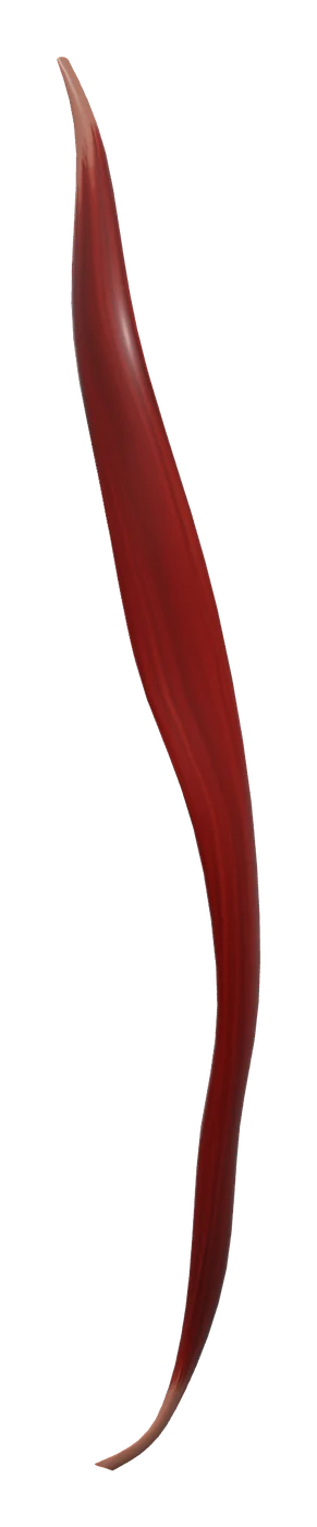
The sartorius, a long strap, crosses the front of the thigh diagonally from the outer side of the hip to the inner side of the knee. It is the longest muscle in the body. Both the quadriceps and the sartorius are supplied by the femoral nerve.
Medial compartment: adductors
The adductor longus, adductor brevis and adductor magnus form layers on the inner thigh, from front to back. The gracilis is a thin strap along the inner edge, the pectineus sits high up near the groin, and the obturator externus lies deep, close to the hip joint. Most of this group is supplied by the obturator nerve. Two exceptions are common: the pectineus usually receives the femoral nerve, and the lower, hamstring part of the adductor magnus is supplied by the sciatic nerve.
Posterior compartment: hamstrings
The biceps femoris lies on the outer side, with a long head from the pelvis and a short head from the femur. The semimembranosus lies on the inner side, under a third hamstring, the semitendinosus, which runs along it. The hamstrings are supplied by the sciatic nerve.
Lateral side
The tensor fasciae latae sits at the front of the hip, on the outer side. It pulls on the iliotibial tract, a thick band of fascia lata that runs down the outside of the thigh to the tibia. It works with the gluteal muscles, which share that band.
What they do together
- The quadriceps extends the knee. It straightens the leg when you stand up from a chair or climb a step. The rectus femoris is the only part that also crosses the hip, so it helps flex the hip too.
- The adductors pull the thigh toward the midline. They keep the legs together, steady the pelvis when you walk and, depending on the position of the hip, help flex or extend it.
- The hamstrings flex the knee and extend the hip. Except for the short head of the biceps femoris, they cross both joints. That is why stretching them is easiest with the knee straight and the hip flexed.
- The sartorius flexes, abducts and laterally rotates the hip and flexes the knee, the movement of sitting cross-legged.
In walking, these groups take turns: the quadriceps takes the load as your heel lands, the hamstrings slow the leg at the end of each swing, and the adductors keep the limb in line under the body.
How to learn them
Start with the compartments and their nerves: femoral in front, obturator on the inner side, sciatic behind. Then learn each compartment from superficial to deep, which is how a dissection would expose them.
- Quadriceps: "one rectus and three vasti". The vasti are named by where they sit: lateral, medial and intermediate.
- Pes anserinus: the sartorius, gracilis and semitendinosus end together on the inner side of the upper tibia. "Say Grace before Tea" (Sartorius, Gracilis, semiTendinosus) gives them in order from front to back.
- Femoral triangle: a classic exam area in the groin, bordered by the inguinal ligament, the sartorius and the adductor longus. From lateral to medial it holds the femoral Nerve, Artery, Vein and Lymphatics, remembered as NAVL.
- Semi-T versus semi-M: the semitendinosus is mostly tendon in its lower half and lies on top; the semimembranosus is broader and lies underneath.
In the app, peel the superficial layer to find the vastus intermedius under the rectus femoris, isolate the adductors to see how they stack, and then run the lower limb quiz: you are given a muscle name and tap it on the 3D body, much like a practical exam. The muscles of the leg are the natural next region.
Frequently asked questions
How many muscles are in the quadriceps?
Four: the rectus femoris and the vastus lateralis, medialis and intermedius. They share one tendon to the patella and the tibia.
What are the hamstring muscles?
The biceps femoris, the semitendinosus and the semimembranosus, at the back of the thigh.
What is the longest muscle in the body?
The sartorius, which runs diagonally across the front of the thigh from the hip bone to the inner side of the tibia.
Which nerve supplies the adductors?
Mainly the obturator nerve. The pectineus often receives the femoral nerve, and the hamstring part of the adductor magnus is supplied by the sciatic nerve.