
Muscle · Lower limb
Semimembranosus: origin, insertion, action and innervation
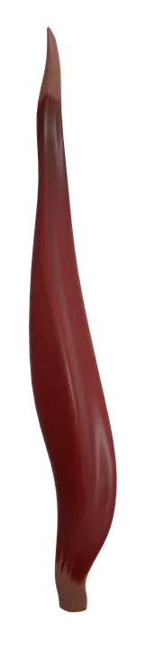
The semimembranosus is a hamstring from the ischial tuberosity to the medial tibial condyle. It extends the hip and flexes the knee (tibial nerve).
Quick answer
- Latin name
- Musculus semimembranosus
- Origin
- Ischial tuberosity of the hip bone
- Insertion
- Back of the medial condyle of the tibia; expansions form the oblique popliteal ligament of the knee
- Action
- Extends the hip; flexes the knee and rotates the leg medially when the knee is bent
- Innervation
- Tibial division of the sciatic nerve (L5–S2)
- Blood supply
- Perforating branches of the deep femoral artery, with branches of the inferior gluteal and popliteal arteries
See it on the 3D body
The semimembranosus lit on the 3D hip and thigh, as in the hip and thigh quiz. Turn the body, peel the layers, tap any structure for its name.
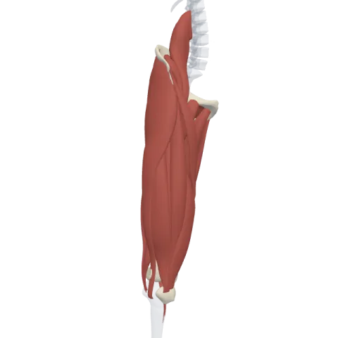
The semimembranosus is one of the three hamstrings, on the medial side of the back of the thigh, running from the ischial tuberosity to the back of the medial condyle of the tibia. It extends the hip, flexes the knee and is supplied by the tibial division of the sciatic nerve (L5–S2).
Where it is
The semimembranosus lies in the posterior compartment of the thigh, on its inner side. It is the deepest and broadest of the medial hamstrings: the semitendinosus runs on top of it, and the two together form the inner wall of the hamstring group. The biceps femoris, the lateral hamstring, runs on the outer side.
Above, its origin is hidden under the gluteus maximus. Below, its tendon forms part of the upper medial border of the popliteal fossa, the diamond-shaped space behind the knee, together with the semitendinosus. See the three compartments together on muscles of the thigh.
Origin and insertion
- Origin: the upper, outer part of the ischial tuberosity of the hip bone, the bony point you sit on. Its origin lies lateral to and deeper than the shared tendon of the semitendinosus and the long head of the biceps femoris.
- Upper tendon: from the ischial tuberosity, the muscle starts as a long, flat, sheet-like tendon. This membrane-like tendon gives the muscle its name.
- Insertion: the back of the medial condyle of the tibia, in a horizontal groove on its posteromedial side. From this tendon, expansions spread out: one runs up and outward behind the knee as the oblique popliteal ligament, which strengthens the back of the joint capsule, and others reach the fascia over the popliteus and the medial meniscus.
Unlike the semitendinosus, it does not join the pes anserinus, the shared tendon of sartorius, gracilis and semitendinosus on the front of the tibia. It stays at the back.
What it does
- Hip extension: because it starts on the pelvis and crosses the back of the hip, it pulls the thigh backward, as in walking, running and rising from a forward bend.
- Knee flexion: it crosses the back of the knee and bends it.
- Medial rotation of the leg: when the knee is bent, it turns the leg inward, working with the semitendinosus. The biceps femoris does the opposite (lateral rotation).
- Tilting the pelvis: with the leg fixed, the hamstrings pull the back of the pelvis down, which helps straighten the trunk after you bend forward.
Because it crosses two joints, it cannot fully extend the hip and fully flex the knee at the same time. This is why a forward bend with straight knees stretches the hamstrings.
Innervation and blood supply
The semimembranosus is supplied by the tibial division of the sciatic nerve (roots L5, S1 and S2), like the semitendinosus and the long head of the biceps femoris. Only the short head of the biceps femoris uses the common fibular division.
Blood comes mainly from the perforating branches of the deep femoral artery, with contributions from the inferior gluteal artery at the top and branches of the popliteal artery at the bottom.
How to remember it
- Semi-membranosus = half-membrane: its upper tendon is a flat sheet. Semi-tendinosus = half-tendon: its lower half is a long, round cord.
- "Membranosus is under": the semimembranosus lies deep to the semitendinosus, which rides on it like a cord on a mat.
- The hamstrings share three features: ischial tuberosity, tibial nerve, two joints (hip and knee). The short head of the biceps femoris breaks all three rules.
- Medial hamstrings rotate the leg medially; the lateral hamstring rotates it laterally.
Frequently asked questions
Is the semimembranosus a hamstring?
Yes. The hamstrings are the semimembranosus, the semitendinosus and the biceps femoris. The semimembranosus and semitendinosus are the medial hamstrings.
What is the difference between the semimembranosus and the semitendinosus?
Both are medial hamstrings with the same origin area, action and nerve. The semimembranosus is deeper and broader, with a flat upper tendon, and inserts on the back of the medial tibial condyle. The semitendinosus lies on top of it, has a long cord-like lower tendon and inserts on the front of the tibia in the pes anserinus.
Where does the semimembranosus insert?
On the back of the medial condyle of the tibia, with expansions that form the oblique popliteal ligament behind the knee.
Which nerve supplies the semimembranosus?
The tibial division of the sciatic nerve, from roots L5 to S2.
