
Muscle · Lower limb
Psoas major: origin, insertion, action and innervation
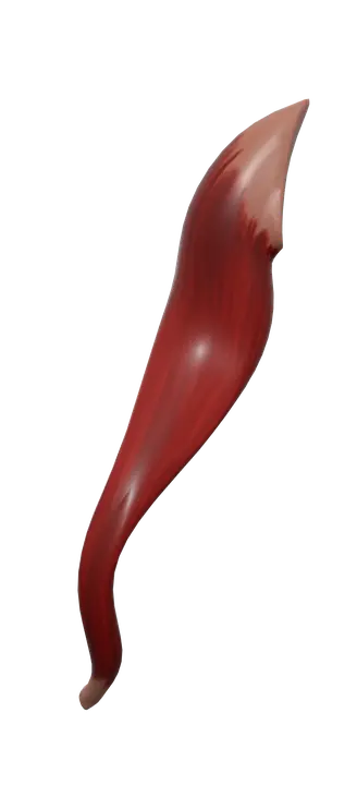
The psoas major is a deep muscle from the T12-L5 vertebrae to the lesser trochanter of the femur. It flexes the hip, with the iliacus (L1-L3 anterior rami).
Quick answer
- Latin name
- Musculus psoas major
- Origin
- Sides of the bodies of T12–L5 and the discs between them; transverse processes of L1–L5
- Insertion
- Lesser trochanter of the femur, with the iliacus (iliopsoas tendon)
- Action
- Flexes the hip; with the thigh fixed, flexes the trunk and bends the lumbar spine to the side
- Innervation
- Anterior rami of the lumbar nerves (L1–L3)
- Blood supply
- Lumbar arteries and the lumbar branch of the iliolumbar artery
See it on the 3D body
The psoas major lit on the 3D pelvis, as in the pelvis quiz. Turn the body, peel the layers, tap any structure for its name.
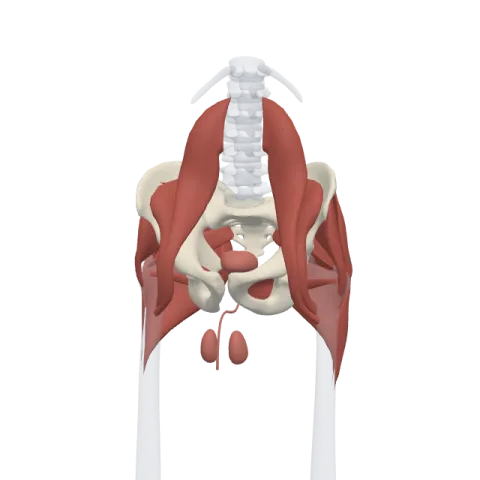
The psoas major is a long, thick muscle on each side of the lumbar spine, running from the T12–L5 vertebrae to the lesser trochanter of the femur. With the iliacus it forms the iliopsoas, the strongest flexor of the hip, and it is supplied by the anterior rami of L1–L3.
Where it is
The psoas major lies deep in the back wall of the abdomen, on either side of the lumbar vertebrae, behind the abdominal organs. From there it runs down along the brim of the pelvis, passes under the inguinal ligament and enters the front of the thigh, where it ends on the inner side of the upper femur.
On its way it is joined by the iliacus, which fills the inner bowl of the ilium. The two share a tendon and are often described together as the iliopsoas. In the upper thigh, the iliopsoas forms part of the floor of the femoral triangle, lateral to the pectineus.
A thin muscle in front of it, the psoas minor, is absent in many people. See the iliopsoas with the gluteal and deep hip muscles on muscles of the hip.
Origin and insertion
- Origin: the sides of the bodies of the T12 to L5 vertebrae and the intervertebral discs between them, and the front and lower borders of the transverse processes of L1 to L5. Between the bodies and the transverse processes there are two layers of the muscle.
- Insertion: the lesser trochanter of the femur, a small bump on the back and inner side of the bone below the neck. The tendon is shared with the iliacus, whose fibers join it on its lateral side.
What it does
- Hip flexion: with the iliacus, it lifts the thigh forward and up, as when you climb stairs, march or kick. It is the main flexor of the hip, helped by the rectus femoris and the sartorius.
- Trunk flexion: when the legs are fixed, it pulls the trunk and pelvis forward, for example when you sit up from lying on your back. The rectus abdominis works with it in this movement.
- Lateral flexion of the lumbar spine: acting on one side, it bends the lumbar spine toward the same side.
- Posture: it helps steady the lumbar spine and pelvis when standing and sitting.
Its role in rotation of the thigh is small and textbooks describe it differently, so exams usually focus on flexion.
Innervation and blood supply
The psoas major is supplied directly by the anterior rami of the lumbar nerves (L1, L2 and L3). The lumbar plexus forms inside the muscle, and several of its branches leave it in a recognizable pattern:
- the femoral nerve comes out of its lateral border;
- the obturator nerve comes out of its medial border;
- the genitofemoral nerve pierces its front surface.
The iliacus, by contrast, is supplied by the femoral nerve. Blood comes from the lumbar arteries and the lumbar branch of the iliolumbar artery.
How to remember it
- Psoas comes from the Greek word for the loin. In beef or pork, the psoas is the tenderloin.
- "Spine to femur": it's the only muscle that connects the lumbar spine directly to the lower limb.
- Psoas + iliacus = iliopsoas, one tendon, one job: hip flexion.
- Lumbar plexus exits: femoral lateral, obturator medial, genitofemoral through the front.
- It starts at T12, the last thoracic vertebra, and covers the whole lumbar spine down to L5.
Frequently asked questions
What is the main function of the psoas major?
It flexes the hip, together with the iliacus. With the thigh fixed, it also flexes the trunk forward.
Where does the psoas major originate and insert?
It originates from the sides of the T12–L5 vertebral bodies and discs and the transverse processes of L1–L5. It inserts on the lesser trochanter of the femur.
What is the difference between the psoas and the iliopsoas?
The psoas major is one muscle. The iliopsoas is the name for the psoas major and the iliacus together, because they share a tendon on the lesser trochanter and the same main action.
Which nerve supplies the psoas major?
The anterior rami of L1 to L3, directly from the lumbar plexus, which forms inside the muscle.
Is the psoas a back muscle or a hip muscle?
It lies in the back wall of the abdomen, next to the lumbar spine, but its main action is on the hip. Anatomy courses usually teach it with the muscles of the hip and the lower limb.
