
Muscle · Lower limb
Tensor fasciae latae: origin, insertion, action and innervation
The tensor fasciae latae runs from the ASIS to the iliotibial tract. It abducts and medially rotates the hip and is supplied by the superior gluteal nerve.
Quick answer
- Latin name
- Musculus tensor fasciae latae
- Origin
- Anterior superior iliac spine and the front of the outer lip of the iliac crest, on the hip bone
- Insertion
- Iliotibial tract, which attaches to the lateral condyle of the tibia
- Action
- Abducts and medially rotates the hip; helps flex it; tenses the fascia lata and iliotibial tract; steadies the extended knee
- Innervation
- Superior gluteal nerve (L4-S1)
- Blood supply
- Ascending branch of the lateral circumflex femoral artery and the superior gluteal artery
See it on the 3D body
The tensor fasciae latae lit on the 3D hip and thigh, as in the hip and thigh quiz. Turn the body, peel the layers, tap any structure for its name.
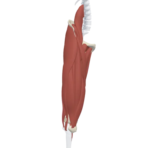
The tensor fasciae latae is a short muscle at the front and side of the hip that runs from the anterior superior iliac spine into the iliotibial tract. It abducts and medially rotates the hip, tightens the fascia lata and is supplied by the superior gluteal nerve (L4-S1).
Where it is
Put your hand on the bony point at the front of your hip, the anterior superior iliac spine (ASIS). Just below and behind it, on the outer side of the thigh, lies the tensor fasciae latae. It sits between two neighbors: the sartorius in front and the gluteus medius behind.
Its name describes its job. The fascia lata is the tough sleeve of deep fascia that wraps the whole thigh. On the outer side, this sleeve thickens into a strong band, the iliotibial tract (IT band), that runs down to the knee. The tensor fasciae latae is the "tensor" (tightener) of this fascia: its fibers end inside the tract, between two layers of fascia.
The gluteus maximus also pulls on the iliotibial tract from behind, so the band is held by one muscle in front and one behind. See both on muscles of the hip.
Origin and insertion
- Origin: the anterior superior iliac spine and the front part of the outer lip of the iliac crest, on the hip bone.
- Insertion: the iliotibial tract, about a third of the way down the thigh. Through the tract, its pull reaches the anterolateral tubercle of the lateral condyle of the tibia (Gerdy's tubercle).
So the muscle itself is short, but its effect travels the length of the thigh through a tendon-like band of fascia.
What it does
- Abduction of the hip: it moves the thigh away from the midline, helping the gluteus medius and minimus.
- Medial rotation of the hip: because it lies in front of the joint's axis, it turns the thigh inward.
- Flexion of the hip: it gives some help in lifting the thigh forward.
- Tensing the fascia lata and iliotibial tract: this makes the outer thigh stiffer and helps steady the knee when it is straight and bearing weight.
During walking it works with the gluteus medius to keep the pelvis level over the standing leg. Lower down, the tract crosses the outer side of the knee, which is why runners and cyclists often talk about the IT band.
Innervation and blood supply
The tensor fasciae latae is supplied by the superior gluteal nerve (L4-S1). This is the same nerve that supplies the gluteus medius and gluteus minimus, which makes sense since the three muscles share the job of abducting and medially rotating the hip.
Its blood comes from the ascending branch of the lateral circumflex femoral artery, with a contribution from the superior gluteal artery.
How to remember it
- Read the name: tensor (tightener) of the fascia lata (broad fascia). The name is the action.
- "Superior trio": the superior gluteal nerve serves the medius, the minimus and the TFL. The inferior gluteal nerve serves the maximus alone.
- A pocket in the hip: find the ASIS with your thumb, slide your fingers down and back, and you are on the TFL. Turn the thigh inward and you feel it tighten.
- Front and back hands on one band: TFL pulls the iliotibial tract from the front, gluteus maximus pulls it from the back.
Test yourself in the hip and thigh quiz.
Frequently asked questions
What does the tensor fasciae latae do?
It abducts and medially rotates the thigh, helps flex the hip, and tightens the fascia lata and iliotibial tract.
What nerve supplies the tensor fasciae latae?
The superior gluteal nerve, from roots L4 to S1.
Where does the tensor fasciae latae insert?
Into the iliotibial tract, which carries its pull down to the lateral condyle of the tibia.
Is the tensor fasciae latae a gluteal muscle?
It is grouped with the gluteal muscles because it shares their nerve and helps them abduct the hip, though it sits at the front of the group.
