
Muscle · Lower limb
Tibialis posterior: origin, insertion, action and innervation
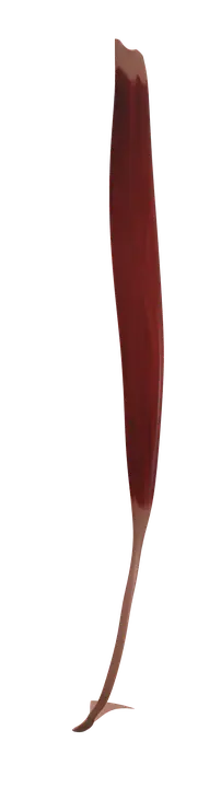
The tibialis posterior is a deep calf muscle from the back of the tibia, fibula and membrane to the navicular. It inverts the foot (tibial nerve).
Quick answer
- Latin name
- Musculus tibialis posterior
- Insertion
- Tuberosity of the navicular and the medial cuneiform; slips to the other cuneiforms, the cuboid, the calcaneus and the bases of metatarsals 2–4
- Action
- Inverts the foot; helps plantar flex the ankle; supports the medial longitudinal arch
- Innervation
- Tibial nerve (L4–L5)
- Blood supply
- Posterior tibial artery, with branches of the fibular artery
See it on the 3D body
The tibialis posterior lit on the 3D leg and foot, as in the leg and foot quiz. Turn the body, peel the layers, tap any structure for its name.
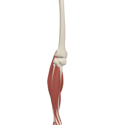
The tibialis posterior is the deepest muscle of the back of the leg, running from the interosseous membrane, tibia and fibula to the navicular bone and the tarsal bones around it. It inverts the foot, supports the medial arch, and is supplied by the tibial nerve (L4–L5).
Where it is
The tibialis posterior lies in the deep part of the posterior compartment of the leg, directly on the interosseous membrane, the sheet of connective tissue that joins the tibia and the fibula. It is covered by the other deep calf muscles and, above them, by the soleus and the gastrocnemius.
It sits between the two long toe flexors: the flexor digitorum longus on the tibial side and the flexor hallucis longus on the fibular side. Above the ankle, its tendon crosses under the flexor digitorum longus to become the most medial of the deep tendons, then runs in a groove behind the medial malleolus, the bony bump on the inner side of the ankle, before passing into the sole. See all the leg compartments on muscles of the leg.
Origin and insertion
- Origin: the back of the interosseous membrane, the lateral part of the posterior surface of the tibia below the soleal line, and the medial part of the posterior surface of the fibula.
- Insertion: the main tendon attaches to the tuberosity of the navicular and the medial cuneiform. From there, slips spread across the underside of the foot to the intermediate and lateral cuneiforms, the cuboid, the sustentaculum tali of the calcaneus and the bases of the second to fourth metatarsals.
This wide, fan-like insertion on the underside of the tarsal bones is what lets it hold up the arch. For the bones themselves, see bones of the foot.
What it does
- Inversion of the foot: it turns the sole of the foot inward. It is the main inverter, working with the tibialis anterior.
- Plantar flexion of the ankle: because its tendon passes behind the medial malleolus, it helps point the foot down, a secondary role next to the soleus and gastrocnemius.
- Supporting the medial longitudinal arch: its tendon pulls up on the navicular, the keystone of the inner arch, and its slips tie the tarsal bones together from below.
- Steadying the foot when walking: it controls how the foot flattens when it takes weight, and helps make it rigid for push-off.
Innervation and blood supply
The tibialis posterior is supplied by the tibial nerve (roots L4 and L5), the nerve of the posterior compartment of the leg, which runs down between the superficial and deep calf muscles.
Blood comes mainly from the posterior tibial artery, which runs with the tibial nerve, with branches of the fibular artery.
How to remember it
- Tibialis posterior = on the back of the tibia. Its partner, the tibialis anterior, is on the front. Both invert the foot; anterior dorsiflexes, posterior plantar flexes.
- "Tom, Dick And Very Nervous Harry": the order of structures behind the medial malleolus, from front to back: Tibialis posterior, flexor Digitorum longus, posterior tibial Artery, Vein, tibial Nerve, flexor Hallucis longus.
- Posterior compartment = tibial nerve = plantar flexion.
- It inserts on the navicular, the keystone of the medial arch: think of it as a sling holding the arch up.
Frequently asked questions
What is the main action of the tibialis posterior?
It inverts the foot. It also helps plantar flex the ankle and holds up the medial longitudinal arch.
Where does the tibialis posterior insert?
Mainly on the tuberosity of the navicular and the medial cuneiform, with slips to the other cuneiforms, the cuboid, the calcaneus and the bases of the second to fourth metatarsals.
What is the difference between the tibialis anterior and the tibialis posterior?
Both invert the foot. The tibialis anterior lies on the front of the leg, dorsiflexes the ankle and is supplied by the deep fibular nerve. The tibialis posterior lies deep in the back of the leg, plantar flexes the ankle and is supplied by the tibial nerve.
Which nerve supplies the tibialis posterior?
The tibial nerve, from roots L4 and L5.
Which tendon lies closest to the medial malleolus?
The tibialis posterior tendon. It runs in a groove right behind the medial malleolus and is the most anterior of the structures passing there, followed by the flexor digitorum longus, the posterior tibial vessels, the tibial nerve and the flexor hallucis longus.
