
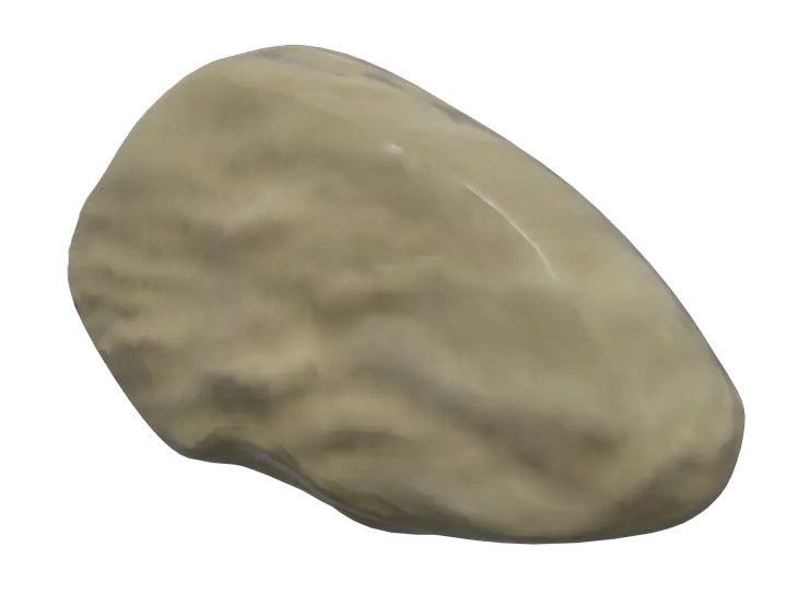
See it on the 3D body
The navicular bone lit on the 3D leg and foot, as in the leg and foot quiz. Turn the body, peel the layers, tap any structure for its name.
Quick answer
- Latin name
- Os naviculare
- Type
- Short bone (tarsal bone)
- Location
- Medial side of the midfoot, in front of the head of the talus
- Joints
- Talonavicular joint with the talus; cuneonavicular joint with the three cuneiform bones; sometimes a small joint with the cuboid
The navicular bone of the foot is a boat-shaped tarsal bone on the inner side of the midfoot, between the head of the talus behind and the three cuneiform bones in front. Its tuberosity, a bump you can feel on the inner side of the foot, receives the main tendon of the tibialis posterior.
Where it is and what it touches
The navicular sits on the medial side of the foot, roughly halfway between the ankle and the base of the big toe. It belongs to the middle group of tarsal bones and links the back of the foot to the front.
- Behind, its concave surface cups the rounded head of the talus at the talonavicular joint. This joint, with the calcaneocuboid joint beside it, forms the transverse tarsal joint, an S-shaped line across the foot.
- In front, it meets the medial, intermediate and lateral cuneiforms at the cuneonavicular joint.
- On the lateral side, it often has a small contact with the cuboid.
It does not touch the calcaneus directly, but a strong ligament runs from the calcaneus to its underside: the plantar calcaneonavicular ligament, or spring ligament, which helps cradle the head of the talus.
The navicular lies near the top of the medial longitudinal arch, the high arch on the inner side of the foot. See where it fits among the 26 bones in bones of the foot.
Shape and landmarks
The name comes from the Latin navicula, a little boat: seen from the side, the bone looks like a small hull, hollow at the back.
- Posterior surface. Oval and concave, covered with cartilage, for the head of the talus.
- Anterior surface. Convex and divided by two faint ridges into three facets, one for each cuneiform.
- Dorsal surface. Rough and convex, for ligaments that tie it to its neighbors.
- Plantar surface. Rough and concave, for the spring ligament and other plantar ligaments.
- Navicular tuberosity. A rounded projection on the medial side, pointing down and inward. You can find it a little in front of and below the medial malleolus, the bump on the inside of the ankle.
Muscles that attach to the navicular
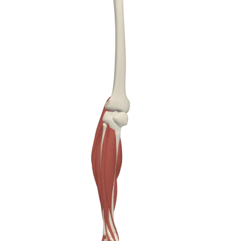
The tibialis posterior is the deepest muscle at the back of the leg. It starts on the tibia, the fibula and the membrane between them, runs behind the medial malleolus, and inserts mainly on the navicular tuberosity. From there, slips spread to the cuneiforms, the cuboid and the bases of the middle metatarsals.
Its pull points the foot down (plantar flexion) and turns the sole inward (inversion). Because its tendon passes under the inner side of the foot, it also supports the medial arch from below while you stand and walk.
The tibialis anterior, at the front of the shin, takes a nearby route but does not insert on the navicular: its tendon passes in front of it to the medial cuneiform and the base of the first metatarsal. Both muscles are part of the muscles of the leg.
What the navicular does
- It transfers weight. Load from the talus passes through the navicular to the cuneiforms and on to the first three metatarsals, toward the ball of the foot.
- It lets the foot twist. The talonavicular joint is one of the most mobile joints of the foot. Together with the subtalar joint, it lets the forefoot turn inward and outward relative to the heel, so the foot can adapt to uneven ground.
- It helps hold the arch. It sits near the top of the medial arch, held from below by the spring ligament and the tibialis posterior.
Navicular of the foot, not of the hand
Older texts and some clinicians use "navicular" for a bone of the wrist too. That wrist bone is now called the scaphoid, one of the eight carpal bones. In current anatomical terminology, "navicular bone" means the bone of the foot.
In Anatomy Atlas 3D
In the app, the bone appears as "Navicular bone" in each foot, with its Latin name, os naviculare. Tap it, then tap the tibialis posterior to follow its tendon behind the ankle and down to the tuberosity. The app is free on iPad and iPhone and works offline.
Frequently asked questions
Where is the navicular bone located?
On the inner side of the midfoot, in front of the head of the talus and behind the three cuneiform bones.
What muscle attaches to the navicular tuberosity?
The tibialis posterior. Its tendon inserts mainly on the tuberosity and sends slips to neighboring tarsal and metatarsal bones.
Is the navicular the same as the scaphoid?
No. The scaphoid is a carpal bone of the wrist that was once also called the navicular. Today the navicular is the tarsal bone of the foot.
How many bones does the navicular articulate with?
Usually four or five: the talus, the three cuneiforms and, in many people, the cuboid.
Why is it called navicular?
From the Latin for "little boat", because of its shape: hollow at the back like the inside of a small hull.
Muscles that insert here
From the anatomical data behind Anatomy Atlas 3D (Z-Anatomy, reviewed). In the app, tap the bone, then each muscle around it to open its card.
