
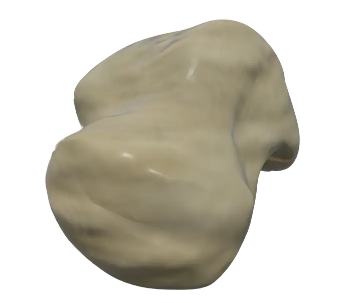
See it on the 3D body
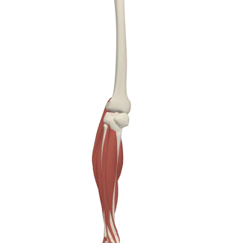
The talus lit on the 3D leg and foot, as in the leg and foot quiz. Turn the body, peel the layers, tap any structure for its name.
Quick answer
- Latin name
- Talus
- Type
- Short bone (tarsal bone)
- Location
- Top of the foot, between the leg bones above and the calcaneus below
The talus is the ankle bone: the only bone of the foot that articulates with the leg, wedged between the tibia and fibula above and the calcaneus below. It passes the whole weight of the body from the leg to the foot, yet no muscle attaches to it.
Where it is and what it touches
The talus is the second largest of the seven tarsal bones, after the calcaneus. It sits at the top of the foot, at the hinge between the leg and the foot.
It has three sets of joints, and much of its surface is covered with cartilage:
- Above and on both sides, with the tibia and the fibula, at the ankle (talocrural) joint. The lower end of the tibia and the two malleoli, the bony bumps on each side of the ankle, wrap around the talus like a mortise around a tenon.
- Below, with the calcaneus, at the subtalar joint.
- In front, with the navicular, at the talonavicular joint.
These three joints are what let the foot move up and down and tilt inward and outward. For the full picture of the 26 bones, see bones of the foot.
Parts and landmarks
Anatomists divide the talus into a head, a neck and a body.
Body
- The trochlea is the rounded, pulley-shaped upper surface. It fits under the tibia and moves forward and back as the foot bends up and down. It is wider in front than behind, which makes the ankle more snug when the foot is pulled up.
- The medial malleolar facet, comma-shaped, faces the medial malleolus of the tibia.
- The lateral malleolar facet, larger and triangular, faces the lateral malleolus of the fibula. Below it, the lateral process juts out to the side.
- The posterior process at the back has a medial and a lateral tubercle, with a groove between them for the tendon of the flexor hallucis longus.
- Underneath, the body carries a large facet for the calcaneus.
Neck
The neck is the narrow part between the body and the head. Its underside is marked by a deep groove, the sulcus tali, which meets the matching groove on the calcaneus to form the tarsal sinus. The neck rests on the sustentaculum tali, a shelf on the inner side of the calcaneus.
Head
The rounded head points forward and slightly inward. It fits into a cup on the back of the navicular. Underneath, it also rests on smaller facets of the calcaneus and on a ligament between the calcaneus and the navicular, the spring ligament, which supports it from below.
Why no muscle attaches to the talus
Most bones have muscles pulling on them, but the talus is an exception: its surface is taken up by joint surfaces and ligaments, and the tendons of the leg muscles run past it to insert farther along the foot. The tibialis anterior crosses in front of it to reach the medial cuneiform and first metatarsal; the calf muscles pass behind it to the calcaneus; the fibular muscles curve around the lateral malleolus to the outer side of the foot.
The talus moves because the bones around it move. When the calf muscles lift the heel, or the muscles in front of the shin raise the foot, the talus rolls within the ankle joint as a passive link.
The anatomical data behind the app agrees: it lists no muscle origin or insertion on the talus.
Movements at its joints
- Ankle joint. A hinge joint. It allows dorsiflexion, raising the top of the foot toward the shin, and plantar flexion, pointing the foot down.
- Subtalar and talonavicular joints. Together they allow inversion, turning the sole inward, and eversion, turning it outward. This helps the foot adapt to uneven ground.
In Anatomy Atlas 3D
In the app, both ankle bones are named "Talus". Tap it and turn the ankle to see the trochlea in its socket, or turn the foot over to see how the head sits on the navicular and the calcaneus. Anatomy Atlas 3D is free on iPad and iPhone, with English and Latin names.
Frequently asked questions
Is the talus the same as the ankle bone?
Yes. "Ankle bone" usually means the talus, although the bumps you feel on each side of the ankle are the malleoli of the tibia and fibula.
Why do no muscles attach to the talus?
Its surface is mostly covered by joint surfaces and ligaments. Tendons pass around it to insert on other bones of the foot, and the talus moves with them as a passive link.
What bones does the talus articulate with?
Four: the tibia and fibula at the ankle joint, the calcaneus at the subtalar joint, and the navicular at the talonavicular joint.
What is the trochlea of the talus?
The pulley-shaped upper surface of the body of the talus, which fits under the tibia in the ankle joint.
Which tarsal bone is the largest, the talus or the calcaneus?
The calcaneus. The talus is the second largest tarsal bone.
