
See them on the 3D body
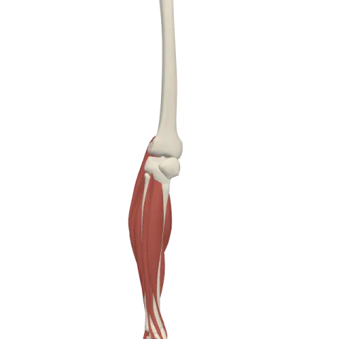
The cuneiform bones (medial, intermediate, lateral) lit on the 3D leg and foot, as in the leg and foot quiz. Turn the body, peel the layers, tap any structure for its name.
Quick answer
- Latin name
- Ossa cuneiformia (mediale, intermedium, laterale)
- Type
- Short bones (three tarsal bones)
- Location
- Midfoot, between the navicular behind and the first three metatarsals in front, beside the cuboid
- Joints
- Cuneonavicular joint with the navicular; intercuneiform joints between the three; cuneocuboid joint with the cuboid; tarsometatarsal joints with the first three metatarsals (the lateral cuneiform also touches the fourth)
The cuneiform bones are three small, wedge-shaped tarsal bones in the middle of the foot: the medial, intermediate and lateral cuneiforms. They sit side by side between the navicular behind and the bases of the first three metatarsals in front, and their wedge shape helps build the transverse arch of the foot.
Where they are and what they touch
"Cuneiform" means wedge-shaped, from the Latin word for wedge. The three bones form a row across the inner half of the midfoot, numbered by position from the big-toe side:
- the medial cuneiform, on the inner edge of the foot, behind the first metatarsal;
- the intermediate cuneiform, in the middle, behind the second metatarsal;
- the lateral cuneiform, behind the third metatarsal, next to the cuboid.
Behind, all three share the front surface of the navicular, which carries three facets, one for each. Together with the navicular and the cuboid they make up the midfoot. The full set of tarsals, from the talus to the cuboid, is on the bones of the foot page.
Medial cuneiform
The medial cuneiform is the largest of the three. Unlike the other two, its wedge is set with the broad edge down, toward the sole, and the thin edge up.
- Behind, it meets the navicular. In front, a large kidney-shaped facet meets the first metatarsal.
- On its outer side it meets the intermediate cuneiform and the side of the base of the second metatarsal.
- Its inner surface is easy to feel on the inner edge of the foot, in front of the bump of the navicular.
Two major tendons reach it from opposite sides. The tibialis anterior comes down the front of the leg and inserts on its inner and lower surface, along with the base of the first metatarsal. The fibularis longus crosses the sole from the outer side and inserts on its lower outer corner, also next to the first metatarsal.
Intermediate cuneiform
The intermediate cuneiform is the smallest of the three. Its wedge points down, with the broad side facing the top of the foot.
- Behind, it meets the navicular; on each side, the medial and lateral cuneiforms.
- In front, it meets only the second metatarsal.
Because it is shorter than its two neighbors, its front surface sits farther back. The medial and lateral cuneiforms project beyond it and create a slot that the base of the second metatarsal fits into. This locks the second metatarsal in place and makes the middle of the foot rigid.
Lateral cuneiform
The lateral cuneiform is medium-sized, between the other two. Like the intermediate cuneiform, its broad side faces up.
- Behind, it meets the navicular. On its inner side, it meets the intermediate cuneiform and the second metatarsal; on its outer side, the cuboid.
- In front, it meets mainly the third metatarsal, with small contacts with the second and fourth.
The short flexor of the big toe, the flexor hallucis brevis, starts in part from its underside and the cuboid.
How the wedges build the arch
Seen in a cross section through the midfoot, the intermediate and lateral cuneiforms are narrow at the sole and wide on top, like the stones of a stone arch. With the medial cuneiform and the cuboid at each end, they form the transverse arch: the foot is curved from side to side, high on the inner side and low on the outer side. Ligaments on the sole and the tendons of the fibularis longus and tibialis posterior hold the wedges together.
Joints
- Cuneonavicular joint: the three cuneiforms against the front of the navicular.
- Intercuneiform joints: between neighboring cuneiforms.
- Cuneocuboid joint: the lateral cuneiform against the cuboid.
- Tarsometatarsal joints: the cuneiforms against the bases of the first three metatarsals.
All of these are flat, gliding joints held by strong ligaments. They move only a little, but together they help the foot change shape as it meets the ground.
Muscles attached
- Tibialis anterior: medial cuneiform and first metatarsal. It lifts the foot and turns the sole inward.
- Fibularis longus: medial cuneiform and first metatarsal, from the outer side. It points the foot and turns the sole outward.
- Tibialis posterior: its main tendon inserts on the navicular, and slips spread to all three cuneiforms.
- Flexor hallucis brevis: starts in part on the lateral cuneiform.
How to remember them
- 1, 2, 3 behind 1, 2, 3. Medial, intermediate and lateral cuneiforms sit behind metatarsals 1, 2 and 3.
- Big, small, medium. Medial is the largest, intermediate the smallest, lateral in between.
- The odd wedge. Only the medial cuneiform has its broad edge on the sole.
- The short one makes the slot. The intermediate cuneiform is set back, so the second metatarsal plugs into the gap.
In Anatomy Atlas 3D
The app shows each cuneiform as a separate structure: "Medial cuneiform bone", "Intermediate cuneiform bone" and "Lateral cuneiform bone". Look at the foot from above to see the slot for the second metatarsal, then from below to see where the tibialis anterior and fibularis longus meet. Try them in the leg and foot quiz. The app is free on iPad and iPhone.
Frequently asked questions
How many cuneiform bones are there?
Three in each foot: medial, intermediate and lateral.
Which cuneiform is the largest?
The medial cuneiform. The intermediate cuneiform is the smallest.
Which muscles attach to the medial cuneiform?
The tibialis anterior on its inner and lower surface and the fibularis longus on its lower outer corner. A slip of the tibialis posterior also reaches it.
What does "cuneiform" mean?
Wedge-shaped. The name comes from the Latin word for a wedge, the same root as cuneiform writing.
Muscles that insert here
From the anatomical data behind Anatomy Atlas 3D (Z-Anatomy, reviewed). In the app, tap the bone, then each muscle around it to open its card.
