
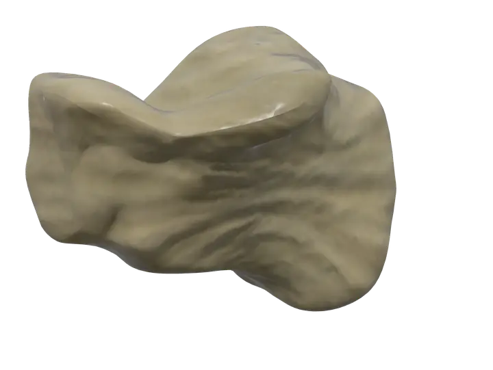
See it on the 3D body
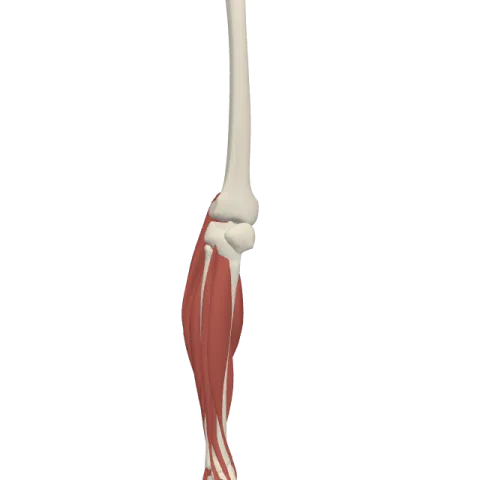
The calcaneus lit on the 3D leg and foot, as in the leg and foot quiz. Turn the body, peel the layers, tap any structure for its name.
Quick answer
- Latin name
- Calcaneus
- Type
- Short bone (tarsal bone)
- Location
- Back of the foot, under the talus, forming the heel
- Joints
- Subtalar (talocalcaneal) joint with the talus; calcaneocuboid joint with the cuboid
The calcaneus is the heel bone: the largest of the seven tarsal bones and the first part of the foot to touch the ground when you walk. It carries the weight passed down from the talus above it and gives the calf muscles a long lever through the Achilles tendon.
Where it is and what it touches
The calcaneus forms the back of the foot and the bony heel you stand on. It lies below the talus and does not touch the tibia or the fibula: the talus sits between them.
It has two main joints:
- Above, with the talus, at the subtalar joint. The two bones meet through several facets, and their movement against each other tilts the sole of the foot inward (inversion) and outward (eversion).
- In front, with the cuboid, at the calcaneocuboid joint. Together with the joint between the talus and the navicular, it forms the transverse tarsal joint across the middle of the foot.
The calcaneus also makes the back pillar of the longitudinal arch of the foot. See how it fits with the other 25 bones in bones of the foot.
Shape and landmarks
The calcaneus is roughly a rectangular block, longer than it is wide, with six surfaces.
- Calcaneal tuberosity. The back end of the bone, the heel itself. Its smooth upper part is separated from the Achilles tendon by a small bursa; the tendon attaches lower down, on the middle of the back surface. Underneath, the tuberosity has a medial process and a lateral process that take the weight of the heel and anchor the sole muscles and the plantar aponeurosis.
- Talar facets. On the upper surface, a large posterior facet and smaller middle and anterior facets articulate with the talus.
- Calcaneal sulcus. A groove between the posterior and middle facets. With a matching groove on the talus, it forms the tarsal sinus, a tunnel you can feel as a soft hollow in front of the lateral ankle bone. Strong ligaments that bind the two bones run in it.
- Sustentaculum tali. A shelf projecting from the medial side. Its upper surface carries the middle facet and supports the neck of the talus, which is where its Latin name, "support of the talus", comes from. The tendon of the flexor hallucis longus runs in a groove beneath it.
- Fibular (peroneal) trochlea. A small ridge on the lateral surface, between the tendons of the fibularis longus and brevis on their way to the foot.
- Cuboid articular surface. The front end, shaped like a saddle, for the cuboid.
Muscles that attach to the calcaneus
Insertions, at the back. The gastrocnemius and the soleus, the two main calf muscles, share one tendon, the calcaneal (Achilles) tendon, the thickest and strongest tendon in the body. It inserts on the back of the calcaneal tuberosity. When the calf contracts, it pulls the heel up and points the foot down, the movement called plantar flexion.
Origins, underneath. Several short muscles of the sole start on the processes of the tuberosity:
- the abductor hallucis, along the medial side of the sole, to the great toe;
- the flexor digitorum brevis, in the middle of the sole, to the lateral four toes;
- the abductor digiti minimi, along the lateral side, to the little toe.
Origins, on top. The extensor digitorum brevis and extensor hallucis brevis, the only muscles on the back of the foot, start on the upper and lateral surface of the front of the calcaneus and fan out to the toes.
What the calcaneus does
- It bears weight. At heel strike, the calcaneus takes the impact first. Its inner structure of spongy bone, with struts lined up along the lines of stress, helps it carry loads several times body weight during running.
- It works as a lever. The tuberosity sticks out behind the ankle, so the calf muscles pull on a long arm. This lets you rise onto your toes and push off when walking.
- It shapes the arch. As the back end of the longitudinal arch, it holds up one side of a curved structure that spreads weight between the heel and the balls of the foot.
In Anatomy Atlas 3D
In the free app for iPad and iPhone, the left and right heel bones appear as "Calcaneus". Tap it to highlight it, then tap the gastrocnemius or the soleus to follow the Achilles tendon down to its insertion, or turn the foot over to see the sole muscles that start there.
Frequently asked questions
Is the calcaneus the largest bone of the foot?
Yes. It is the largest tarsal bone and the largest bone of the foot.
What attaches to the back of the heel bone?
The calcaneal (Achilles) tendon, shared by the gastrocnemius and the soleus. It attaches to the back of the calcaneal tuberosity.
What is the sustentaculum tali?
A shelf of bone on the medial side of the calcaneus that supports the talus. The tendon of the flexor hallucis longus passes beneath it.
Which bones does the calcaneus articulate with?
Two: the talus above, at the subtalar joint, and the cuboid in front, at the calcaneocuboid joint.
How many tarsal bones are there?
Seven: talus, calcaneus, navicular, cuboid and the three cuneiforms.
Muscles that originate here
Muscles that insert here
From the anatomical data behind Anatomy Atlas 3D (Z-Anatomy, reviewed). In the app, tap the bone, then each muscle around it to open its card.
