
Muscle · Lower limb
Fibularis longus: origin, insertion, action and innervation
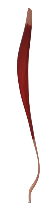
The fibularis longus runs from the upper lateral fibula to the first metatarsal and medial cuneiform, everts the foot, and has the superficial fibular nerve.
Quick answer
- Latin name
- Musculus fibularis longus
- Origin
- Head and upper two-thirds of the lateral surface of the fibula
- Insertion
- Base of the first metatarsal and the medial cuneiform, on their plantar and lateral sides
- Action
- Everts the foot; helps plantar flex the ankle; supports the arches of the foot
- Innervation
- Superficial fibular nerve (L5–S1)
- Blood supply
- Branches of the anterior tibial and fibular arteries
See it on the 3D body
The fibularis longus lit on the 3D leg and foot, as in the leg and foot quiz. Turn the body, peel the layers, tap any structure for its name.
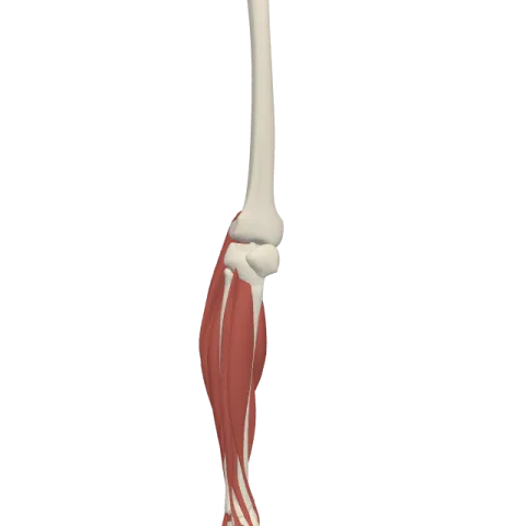
The fibularis longus, also called the peroneus longus, is the long, superficial muscle on the outer side of the leg, running from the upper fibula to the base of the first metatarsal and the medial cuneiform. It everts the foot, helps plantar flex the ankle, and is supplied by the superficial fibular nerve (L5–S1).
Where it is
The fibularis longus lies in the lateral compartment of the leg, on the outer side of the fibula, just under the skin. It covers the shorter fibularis brevis, which shares the compartment with it. In front of it, across a sheet of fascia, is the anterior compartment with the tibialis anterior; behind it is the posterior compartment with the soleus and the other calf muscles.
Its long tendon has an unusual route. It runs down behind the lateral malleolus, the bony bump on the outside of the ankle, then along the outer side of the calcaneus, and finally turns into the sole through a groove on the cuboid bone. From there it crosses the whole width of the sole to the inner side of the foot. When you turn the sole of the foot outward, its tendon stands out as a cord just behind the outer ankle bone. See all three compartments of the leg on muscles of the leg.
Origin and insertion
- Origin: the head of the fibula and the upper two-thirds of the lateral surface of the fibula, with some fibers from the lateral condyle of the tibia and the surrounding fascia and intermuscular septa.
- Insertion: after crossing the sole, the tendon attaches to the lateral and plantar side of the base of the first metatarsal and the medial cuneiform.
The tibialis anterior inserts on the same two bones, from the inner side. The two tendons together form a "stirrup" under the middle of the foot. For the bones involved, see bones of the foot.
What it does
- Eversion of the foot: it turns the sole of the foot outward. It is the strongest everter, with the fibularis brevis.
- Plantar flexion of the ankle: because its tendon passes behind the lateral malleolus, it helps point the foot down, as when you rise onto your toes. This is a weaker, helping role compared with the gastrocnemius and soleus.
- Supporting the arches: its tendon, running across the sole, helps maintain the lateral longitudinal arch and the transverse arch of the foot.
- Balance on uneven ground: it steadies the foot and leg when you stand on one leg, walk on a slope or land from a jump.
Innervation and blood supply
The fibularis longus is supplied by the superficial fibular nerve (roots L5 and S1), the nerve of the lateral compartment. It branches from the common fibular nerve, which winds around the neck of the fibula just below the knee. The other branch, the deep fibular nerve, supplies the anterior compartment instead, including the tibialis anterior.
Blood comes from branches of the anterior tibial artery and the fibular artery.
How to remember it
- Fibularis = on the fibula. The older name peroneus comes from peronē, the Greek word for the same bone (both words originally meant a pin or brooch), so the two names mean the same thing.
- "Lateral compartment, superficial fibular nerve, eversion": the three facts go together for both fibularis longus and brevis.
- Longus goes long: its tendon travels the farthest, all the way across the sole. Brevis stops short, on the fifth metatarsal.
- The stirrup: tibialis anterior on the inside, fibularis longus on the outside, both ending on the medial cuneiform and first metatarsal.
Frequently asked questions
What is the difference between the fibularis longus and the peroneus longus?
There is none. They are two names for the same muscle. Fibularis is the current international term; peroneus is the older name still used in many clinics and textbooks.
What is the main action of the fibularis longus?
It everts the foot, turning the sole outward. It also helps plantar flex the ankle and supports the arches of the foot.
Where does the fibularis longus insert?
On the base of the first metatarsal and the medial cuneiform, on the inner side of the sole, after its tendon has crossed the foot.
Which nerve supplies the fibularis longus?
The superficial fibular nerve, from roots L5 and S1.
