
See them on the 3D body
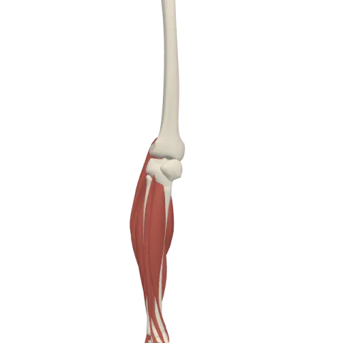
The metatarsal bones (1 to 5) lit on the 3D leg and foot, as in the leg and foot quiz. Turn the body, peel the layers, tap any structure for its name.
Quick answer
- Latin name
- Ossa metatarsi
- Type
- Long bones (five metatarsals)
- Location
- Forefoot, between the tarsal bones (cuneiforms and cuboid) and the proximal phalanges of the toes
- Joints
- Tarsometatarsal joints with the three cuneiform bones and the cuboid; intermetatarsal joints between the bases; metatarsophalangeal joints with the phalanges of the toes
The metatarsal bones are the five long bones of the forefoot, numbered 1 to 5 from the big toe to the little toe. They link the tarsal bones to the toes, and their rounded heads form the ball of the foot, which takes the body's weight when you push off to walk.
Where they are and what they touch
The metatarsals lie side by side across the middle and front of the foot. Behind, their bases join the three cuneiform bones and the cuboid. In front, their heads join the proximal phalanges of the toes.
Like the metacarpals of the hand, each one has a base, a shaft and a head. The bases are wedge-shaped and fit together into an arch that is high on the inner side of the foot and low on the outer side. This arch runs across the foot, and the metatarsals also form the front part of the long arches. All 26 foot bones are presented together on the bones of the foot page.
The metatarsals one by one
First metatarsal (big toe)
The first metatarsal is the shortest, thickest and strongest. It carries a large share of the load at push-off.
- Its base meets the medial cuneiform. A rough tuberosity on the underside of the base receives the fibularis longus, and the tibialis anterior inserts on the inner side of the same base.
- Under its head are two grooves for two sesamoid bones, small bones inside the tendons of the flexor hallucis brevis. They protect the tendon of the long flexor of the big toe and lift the head off the ground.
- Its base does not usually join the second metatarsal, so it moves a little on its own.
Second metatarsal
The second metatarsal is usually the longest. Its base is set back, wedged between the medial and lateral cuneiforms, against the short intermediate cuneiform. This keystone position locks it in place, so it is the stiffest of the five.
Third metatarsal
The third metatarsal meets the lateral cuneiform and its two neighbors. It looks much like the second, only a little shorter.
Fourth metatarsal
The fourth metatarsal meets the cuboid and, at the side, the lateral cuneiform. It is smaller than the third.
Fifth metatarsal (little toe)
The fifth metatarsal meets the cuboid. On the outer side of its base is a large bump, the tuberosity of the fifth metatarsal (also called its styloid process). You can feel it halfway along the outer edge of your foot.
- The fibularis brevis inserts on this tuberosity and turns the sole outward.
- The fibularis tertius, when present, inserts on the back of the base and shaft.
- The abductor and the short flexor of the little toe also use the base.
A Jones fracture is a fracture of the base of the fifth metatarsal, just beyond the tuberosity.
Joints
- Tarsometatarsal joints: between the bases of the metatarsals and the cuneiforms and cuboid. They are flat, firm joints with little movement, especially at the second metatarsal. The first and the fourth and fifth move a little more.
- Intermetatarsal joints: small joints between the bases of metatarsals 2 to 5.
- Metatarsophalangeal (MTP) joints: between each head and the proximal phalanx of a toe. They let the toes bend, lift and spread a little. When you rise on your toes, these joints bend upward and the heads take the weight.
Muscles attached
- The fibularis longus crosses the sole and inserts on the base of the first metatarsal and the medial cuneiform.
- The tibialis anterior inserts on the same two bones from the inner side.
- The fibularis brevis inserts on the tuberosity of the fifth metatarsal.
- The tibialis posterior sends small slips to the bases of the second to fourth metatarsals.
- The adductor hallucis starts in part on the bases of the second to fourth metatarsals.
- The dorsal and plantar interossei fill the spaces between the shafts and move the toes.
How to remember them
- Thick one, long two, bump five. The first is the thickest, the second the longest, the fifth has the bump.
- Two tendons, one spot. The fibularis longus and the tibialis anterior meet at the base of the first metatarsal and the medial cuneiform, from opposite sides, like the two ends of a stirrup.
- Feel the bump. Run a finger along the outer edge of your foot from the little toe toward the heel: the bony knob halfway is the fifth metatarsal tuberosity.
- Keystone two. The set-back base of the second metatarsal is the key that locks the middle of the foot.
In Anatomy Atlas 3D
The app names each metatarsal, from "First metatarsal bone" to "Fifth metatarsal bone". Show the tendons of the leg muscles and follow the fibularis longus under the foot to the first metatarsal. Check yourself in the leg and foot quiz. Anatomy Atlas 3D is free on iPad and iPhone.
Frequently asked questions
How many metatarsal bones are in the foot?
Five in each foot, numbered from the big toe (1) to the little toe (5).
Which metatarsal is the longest?
Usually the second. The first is the shortest and thickest.
What is the bump on the outer side of the foot?
The tuberosity of the fifth metatarsal, where the fibularis brevis tendon inserts.
What are the sesamoid bones of the foot?
Two small bones under the head of the first metatarsal, inside the tendons of the flexor hallucis brevis.
Muscles that insert here
From the anatomical data behind Anatomy Atlas 3D (Z-Anatomy, reviewed). In the app, tap the bone, then each muscle around it to open its card.
