
Muscle · Lower limb
Piriformis muscle: origin, insertion, action and innervation
The piriformis is a deep buttock muscle from the front of the sacrum to the greater trochanter. It rotates the extended hip laterally (S1-S2).
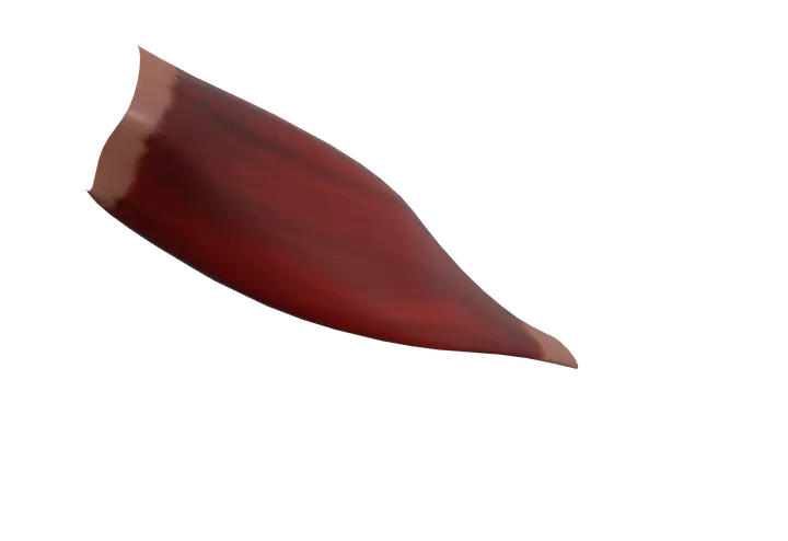
Quick answer
- Latin name
- Musculus piriformis
- Origin
- Front (pelvic) surface of the sacrum, edge of the greater sciatic notch of the hip bone, and the sacrotuberous ligament
- Insertion
- Upper border of the greater trochanter of the femur
- Action
- Rotates the extended hip laterally; abducts the flexed hip; helps hold the head of the femur in the acetabulum
- Innervation
- Nerve to piriformis, from the sacral plexus (S1–S2)
- Blood supply
- Superior and inferior gluteal arteries
See it on the 3D body
The piriformis lit on the 3D hip and thigh, as in the hip and thigh quiz. Turn the body, peel the layers, tap any structure for its name.
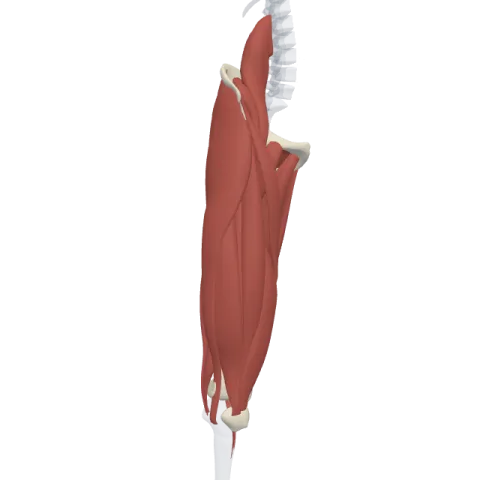
The piriformis is a flat, pear-shaped muscle deep in the buttock, running from the front of the sacrum, through the greater sciatic foramen, to the top of the greater trochanter of the femur. It rotates the extended hip laterally and is supplied by the nerve to piriformis (S1–S2), a branch of the sacral plexus.
Where it is
The piriformis lies deep to the gluteus maximus, below the gluteus medius and gluteus minimus. It is the uppermost of the deep lateral rotators of the hip, a group of small muscles that also includes the superior gemellus, obturator internus, inferior gemellus, quadratus femoris and obturator externus.
It is unusual because part of it lies inside the pelvis: it starts on the inner surface of the sacrum, on the back wall of the pelvic cavity, then leaves the pelvis through the greater sciatic foramen to reach the buttock. Find it with the rest of the deep hip muscles on muscles of the hip.
Origin and insertion
- Origin: the front (pelvic) surface of the sacrum, mainly between and around the second to fourth anterior sacral foramina. Some fibers also come from the upper edge of the greater sciatic notch of the hip bone and from the sacrotuberous ligament.
- Insertion: a rounded tendon on the upper border of the greater trochanter of the femur, often blending with the common tendon of the obturator internus and gemelli.
What it does
- Lateral rotation of the extended hip: when the hip is straight, as in standing, it turns the thigh outward so the toes point away from the midline.
- Abduction of the flexed hip: when the hip is bent to about 90°, as in sitting, its line of pull changes and it moves the thigh outward instead. This change of action with hip position is a classic exam question.
- Stabilizing the hip joint: with the other deep rotators, it works like a rotator cuff of the hip, holding the head of the femur in the acetabulum during walking and standing.
Innervation and blood supply
The piriformis is supplied by the nerve to piriformis, small branches from the anterior rami of S1 and S2 that enter the muscle on its pelvic surface, before it leaves the pelvis.
Blood comes from the superior and inferior gluteal arteries, which pass above and below it, with small branches of the lateral sacral artery.
Why it's a landmark
The piriformis fills most of the greater sciatic foramen and divides it into two passages. Learning what passes above and below it organizes the whole gluteal region:
- Above the piriformis: the superior gluteal nerve, artery and vein, which supply the gluteus medius, gluteus minimus and tensor fasciae latae.
- Below the piriformis: the sciatic nerve, the inferior gluteal nerve and vessels (to the gluteus maximus), the pudendal nerve and internal pudendal vessels, the posterior femoral cutaneous nerve, and the nerves to the obturator internus and quadratus femoris.
In most people, the sciatic nerve passes below the piriformis. In a minority, part or all of the nerve passes through or above the muscle, a normal anatomical variation.
How to remember it
- Piriformis = pear-shaped (Latin pirum, pear): broad at the sacrum, narrow at the trochanter.
- "Superior goes superior": the superior gluteal nerve and vessels pass above the piriformis, the inferior ones below.
- "Rotates when straight, abducts when bent": lateral rotator in extension, abductor in flexion.
- It's the highest of the deep lateral rotators, followed from top to bottom by the superior gemellus, obturator internus, inferior gemellus and quadratus femoris.
Frequently asked questions
What does the piriformis muscle do?
It rotates the hip laterally when the hip is extended, abducts the thigh when the hip is flexed, and helps keep the head of the femur in its socket.
Where is the piriformis located?
Deep in the buttock, under the gluteus maximus. It runs from the front of the sacrum, inside the pelvis, through the greater sciatic foramen to the top of the greater trochanter.
What is the relationship between the piriformis and the sciatic nerve?
The sciatic nerve usually leaves the pelvis just below the piriformis. In some people, part or all of the nerve passes through or above the muscle instead.
Which nerve supplies the piriformis?
The nerve to piriformis, from the anterior rami of S1 and S2 in the sacral plexus.
Why is the piriformis called the key to the gluteal region?
Because every nerve and vessel that leaves the pelvis through the greater sciatic foramen is described by its position above or below the piriformis.
