
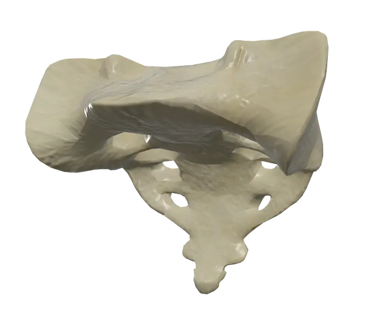
See it on the 3D body
The sacrum lit on the 3D pelvis, as in the pelvis quiz. Turn the body, peel the layers, tap any structure for its name.
Quick answer
- Latin name
- Os sacrum
- Type
- Irregular bone, formed by five fused vertebrae
- Location
- Base of the spine, at the back of the pelvis, between the two hip bones
- Joints
- Lumbosacral joint with the fifth lumbar vertebra; sacro-iliac joints with the two hip bones; sacrococcygeal joint with the coccyx
The sacrum is the triangular bone at the base of the spine, formed by five sacral vertebrae (S1 to S5) that fuse into one bone in early adulthood. Wedged between the two hip bones, it carries the weight of the upper body into the pelvis and the legs.
Where it is and what it touches
The sacrum sits below the fifth lumbar vertebra and above the coccyx, the tailbone. It forms the back wall of the pelvis. Its top is tilted forward, and its front surface curves, so the bone faces partly down and forward.
It has three sets of joints:
- Above, with the fifth lumbar vertebra (L5) at the lumbosacral joint: an intervertebral disc between the bodies, and two small facet joints behind.
- On each side, with the ilium of a hip bone at the sacro-iliac joint. These joints move very little; they are held by some of the strongest ligaments in the body.
- Below, with the coccyx at the sacrococcygeal joint.
The two hip bones, the sacrum and the coccyx together form the bony pelvis. The bones of the spine page shows how the sacrum ends the vertebral column.
Landmarks
Base and apex
- The base is the broad upper surface of S1. Its body meets L5 through the last intervertebral disc of the column.
- The sacral promontory is the front edge of the body of S1, which bulges into the pelvis. It is a reference point for measuring the pelvic inlet.
- The alae, or wings, spread out on each side of the base.
- The superior articular processes rise behind the base to meet L5.
- The apex is the narrow lower end, which joins the coccyx.
Pelvic (front) surface
Smooth and concave. Four transverse ridges mark where the bodies of the five vertebrae fused. At the ends of each ridge, four pairs of anterior sacral foramina let the front branches of the sacral spinal nerves out toward the pelvis and the legs.
Dorsal (back) surface
Rough and convex, with three sets of crests that are the fused remains of vertebral processes:
- the median sacral crest, in the midline, from the spinous processes;
- the intermediate sacral crests, from the articular processes;
- the lateral sacral crests, from the transverse processes.
Four pairs of posterior sacral foramina sit between them for the back branches of the sacral nerves.
Sacral canal and hiatus
The vertebral canal continues down through the sacrum as the sacral canal. At the lower end, the back wall is missing: this opening is the sacral hiatus, flanked by two small horns, the sacral cornua.
Lateral surface
Each side has an ear-shaped auricular surface, covered with cartilage, that meets the ilium at the sacro-iliac joint. Behind it, the rough sacral tuberosity anchors the ligaments that hold the joint.
Muscles that attach to the sacrum
The main muscles attached to the sacrum all start on it:
- The piriformis starts on the pelvic surface, between the anterior foramina. It passes out through the greater sciatic foramen to the greater trochanter of the femur and laterally rotates the hip.
- The gluteus maximus starts partly on the back of the sacrum and the coccyx, as well as on the ilium, then runs to the femur and the iliotibial tract.
- The erector spinae, here the iliocostalis and longissimus, start on the back of the sacrum and run up the spine.
- The latissimus dorsi reaches the sacrum through the thoracolumbar fascia, a broad sheet of connective tissue over the lower back.
Differences between males and females
On average, the female sacrum is shorter, wider and less curved than the male sacrum. This is one of the features that make the female pelvis wider, with a larger inlet and outlet. These are average differences, with plenty of overlap.
What the sacrum does
- It transfers weight from the vertebral column to the hip bones, and from there to the legs, through the sacro-iliac joints.
- It closes the pelvis at the back, protecting pelvic organs.
- It gives exits to nerves: the sacral spinal nerves leave through its foramina to supply the pelvis, the buttocks and much of the leg.
- It anchors muscles of the back and hip.
In Anatomy Atlas 3D
In the app the sacrum is one structure, "Sacrum", with its Latin name, os sacrum. Tap it, then tap the piriformis or the gluteus maximus to see where they start. The app is free on iPad and iPhone and needs no account.
Frequently asked questions
How many vertebrae fuse to form the sacrum?
Five, the sacral vertebrae S1 to S5. They start separate in childhood and fuse during adolescence and early adulthood.
Is the sacrum part of the spine or the pelvis?
Both. It is the lowest fused part of the vertebral column and also forms the back of the pelvis with the coccyx.
What is the sacral promontory?
The front edge of the top of the first sacral vertebra, which projects into the pelvis.
What joints does the sacrum form?
The lumbosacral joint with L5 above, the two sacro-iliac joints with the hip bones, and the sacrococcygeal joint with the coccyx below.
What muscles attach to the sacrum?
The piriformis on the front; the gluteus maximus, the iliocostalis and the longissimus on the back; and the latissimus dorsi through the thoracolumbar fascia.
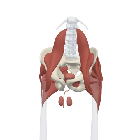
Muscles that originate here
From the anatomical data behind Anatomy Atlas 3D (Z-Anatomy, reviewed). In the app, tap the bone, then each muscle around it to open its card.
