
Muscle · Lower limb
Vastus lateralis: origin, insertion, action and innervation
The vastus lateralis is the largest quadriceps muscle, from the greater trochanter and linea aspera to the patella and tibial tuberosity. It extends the knee.
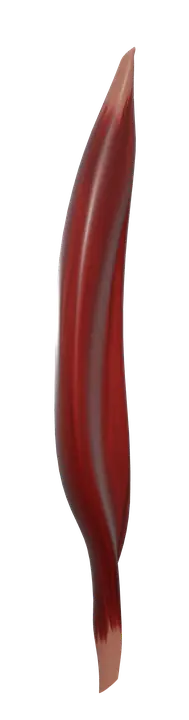
Quick answer
- Latin name
- Musculus vastus lateralis
- Origin
- Greater trochanter, upper intertrochanteric line, gluteal tuberosity and lateral lip of the linea aspera of the femur; lateral intermuscular septum
- Insertion
- Lateral border of the patella through the quadriceps tendon, then the tibial tuberosity of the tibia through the patellar ligament
- Action
- Extends the knee; helps steady the patella
- Innervation
- Femoral nerve (L2–L4)
- Blood supply
- Descending branch of the lateral circumflex femoral artery, and perforating branches of the deep femoral artery
See it on the 3D body
The vastus lateralis lit on the 3D hip and thigh, as in the hip and thigh quiz. Turn the body, peel the layers, tap any structure for its name.
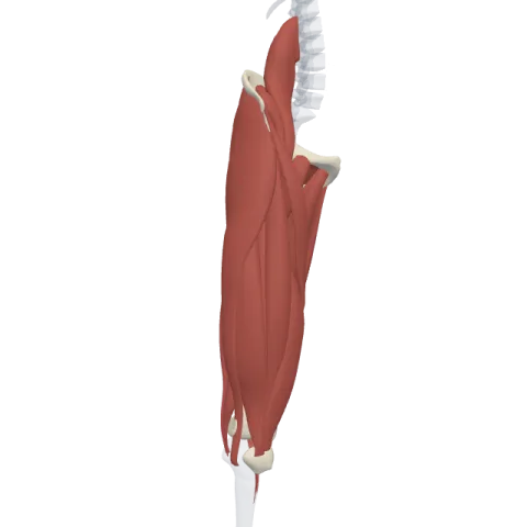
The vastus lateralis is the largest of the four quadriceps muscles, on the outer side of the thigh, running from the upper femur to the patella. It extends the knee and is supplied by the femoral nerve (L2–L4).
Where it is
The vastus lateralis forms most of the bulk on the lateral side of the thigh, between the hip and the knee. It wraps around the outer side of the femur, from the front to the back, and sits next to the rectus femoris, which lies in front of it in the middle of the thigh.
Its surface is covered by the iliotibial tract, the thick band of fascia that runs down the outside of the thigh from the hip to the tibia. Deep to it lies the vastus intermedius, which covers the front of the femoral shaft. On the inner side of the thigh, its partner is the vastus medialis.
Together, rectus femoris, vastus lateralis, vastus intermedius and vastus medialis form the quadriceps femoris, the main muscle group of the anterior compartment of the thigh. See how they fit with the adductors and hamstrings on muscles of the thigh.
Origin and insertion
The vastus lateralis has a long, curved origin that follows the upper and lateral parts of the femur:
- Origin: the upper part of the intertrochanteric line, the front and lower borders of the greater trochanter, the lateral lip of the gluteal tuberosity, and the upper half of the lateral lip of the linea aspera, the rough ridge on the back of the femur. Some fibers also come from the lateral intermuscular septum, the sheet of fascia that separates it from the hamstrings.
- Insertion: its fibers end in a flat tendon that joins the quadriceps tendon and attaches to the lateral border of the patella. From there, the pull reaches the tibial tuberosity of the tibia through the patellar ligament. Some fibers spread into the lateral patellar retinaculum, beside the kneecap.
Because the four quadriceps muscles share this common tendon, the patella sits inside it like a pulley, increasing their leverage on the knee.
What it does
- Knee extension: the vastus lateralis straightens the knee, as when you stand up from a chair, climb stairs, kick a ball or land from a jump. It does not cross the hip, so unlike the rectus femoris, it has no action at the hip joint.
- Steadying the patella: its pull on the lateral side of the kneecap is balanced by the vastus medialis on the medial side, which keeps the patella tracking in the groove at the lower end of the femur.
- Controlling knee bending: when you walk downhill or lower yourself into a squat, it works while lengthening, to control how fast the knee bends.
Innervation and blood supply
Like all the quadriceps, the vastus lateralis is supplied by the femoral nerve (roots L2, L3 and L4), which enters the thigh under the inguinal ligament and splits into branches for the anterior compartment.
Its main blood supply is the descending branch of the lateral circumflex femoral artery, which runs down along its front edge. The perforating branches of the deep femoral artery add to it from behind.
How to remember it
- Vastus = vast, large. Lateralis = on the outer side. It's the largest of the three vasti.
- The quadriceps has four heads, one tendon, one nerve: rectus femoris and the three vasti all end on the patella and all have the femoral nerve.
- "Femoral nerve, front of the thigh": the anterior compartment of the thigh extends the knee and is supplied by the femoral nerve; the medial compartment adducts and has the obturator nerve; the posterior compartment flexes the knee and has the sciatic nerve.
- Only the rectus femoris crosses the hip, so the vasti are pure knee extensors.
Frequently asked questions
What is the main action of the vastus lateralis?
It extends the knee. It works with the other three quadriceps muscles and does not move the hip.
Is the vastus lateralis part of the quadriceps?
Yes. The quadriceps femoris is made of the rectus femoris, vastus lateralis, vastus intermedius and vastus medialis. The vastus lateralis is the largest of the four.
Where does the vastus lateralis originate and insert?
It originates from the greater trochanter, the intertrochanteric line, the gluteal tuberosity and the linea aspera of the femur. It inserts on the patella through the quadriceps tendon and on the tibial tuberosity through the patellar ligament.
Which nerve supplies the vastus lateralis?
The femoral nerve, from roots L2 to L4, which supplies all four quadriceps muscles.
Why do nursing students learn the vastus lateralis?
It is a large, thick muscle on the outer thigh with few major vessels or nerves on its surface, so it is one of the standard sites taught for intramuscular injections, especially in infants. Knowing its position relative to the femur and the knee is part of learning that landmark.
