
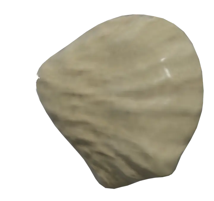
See it on the 3D body
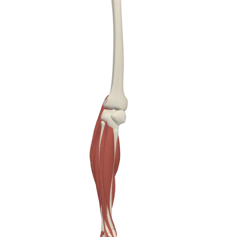
The patella lit on the 3D leg and foot, as in the leg and foot quiz. Turn the body, peel the layers, tap any structure for its name.
Quick answer
- Latin name
- Patella
- Type
- Sesamoid bone
- Location
- Front of the knee, inside the tendon of the quadriceps
The patella, or kneecap, is a small triangular bone at the front of the knee, and the largest sesamoid bone in the body: it grows inside the tendon of the quadriceps. It glides in a groove on the lower end of the femur and acts as a pulley that makes the quadriceps more effective at straightening the knee.
Where it is and what it touches
The patella sits in front of the knee joint, just under the skin. You can feel its whole outline when the leg is straight and relaxed, and even push it gently from side to side, because the quadriceps is slack in that position.
It has only one bony partner. Its back surface rests against the patellar surface of the femur, a smooth groove between the two femoral condyles. This contact forms the patellofemoral joint, one of the three articulations of the knee joint.
The patella does not touch the tibia. Instead, a thick band, the patellar ligament, runs from its lower tip down to the tibial tuberosity, the bump at the top of the shin. The tendon of the quadriceps above, the patella in the middle and the patellar ligament below form one continuous line of pull from the thigh to the leg.
Shape and landmarks
- The base is the broad upper edge. The quadriceps tendon attaches to it.
- The apex is the pointed lower end. The patellar ligament starts there.
- The anterior surface is rough and slightly convex, crossed by tendon fibers.
- The articular surface on the back is covered with thick cartilage. A vertical ridge splits it into a lateral facet, usually the larger one, and a medial facet. The ridge fits into the groove of the femur and keeps the patella on track as the knee bends.
- The medial and lateral borders receive fibers from the vastus medialis and vastus lateralis, which also help guide the bone.
Because it grows in a tendon, the patella is held in place by soft tissue rather than by other bones: the quadriceps above, the patellar ligament below, and thin bands of tissue on each side called retinacula.
Muscles that attach to the patella
All four parts of the quadriceps femoris, the large muscle group on the front of the thigh, converge on the patella:
- The rectus femoris, the only part that also crosses the hip, inserts on the base through the quadriceps tendon.
- The vastus intermedius, deep to the rectus femoris, joins the same tendon.
- The vastus lateralis and vastus medialis insert on the base and along the lateral and medial borders.
From the patella, their pull continues through the patellar ligament to the tibia. That is why anatomy texts often say the quadriceps "inserts on the tibial tuberosity via the patella": both descriptions refer to the same pathway. You can see the four heads and how they meet in the muscles of the thigh.
What the patella does
- It improves leverage. By lifting the quadriceps tendon away from the center of the knee, the patella increases the distance between the line of pull and the axis of the joint. The same muscle force produces more turning effect, which helps when you stand up from a chair, climb stairs or jump.
- It reduces friction. Without the patella, the quadriceps tendon would rub directly on the femur. The smooth cartilage of the patella spreads the load over a wider surface.
- It protects the front of the knee. When the knee is bent, the patella covers the front of the femoral condyles.
As the knee bends from straight to fully flexed, the patella slides down and slightly sideways in the femoral groove, and the area of contact on its back surface shifts from the lower part to the upper part of the facets.
How it develops
At birth the patella is made of cartilage. It usually starts to ossify in early childhood, from one or more centers, and becomes fully bony during childhood and adolescence. Babies do have kneecaps; they just don't show clearly on X-rays yet.
In Anatomy Atlas 3D
The app shows both patellae as one named structure, "Patella", in front of the knee. Tap it, then turn the model to see the facets on its back surface and the quadriceps muscles that converge on it. The app is free on iPad and iPhone, works offline and needs no account.
Frequently asked questions
Why is the patella called a sesamoid bone?
Because it develops inside a tendon, the quadriceps tendon, like the small sesame-seed-shaped bones found in some tendons of the hand and foot. It is the largest bone of this type.
Is the patella part of the knee joint?
Yes. Its back surface articulates with the femur in the patellofemoral joint, which shares the same joint cavity as the joint between the femur and the tibia.
What muscles attach to the kneecap?
The four parts of the quadriceps: rectus femoris, vastus intermedius, vastus lateralis and vastus medialis. Their tendon attaches to the base and borders of the patella and continues as the patellar ligament to the tibia.
Does the patella connect to the tibia?
Not directly. The patellar ligament links the apex of the patella to the tibial tuberosity.
Which facet of the patella is larger?
Usually the lateral facet. It matches the lateral side of the femoral groove, which is higher and broader than the medial side.
Muscles that insert here
From the anatomical data behind Anatomy Atlas 3D (Z-Anatomy, reviewed). In the app, tap the bone, then each muscle around it to open its card.
