
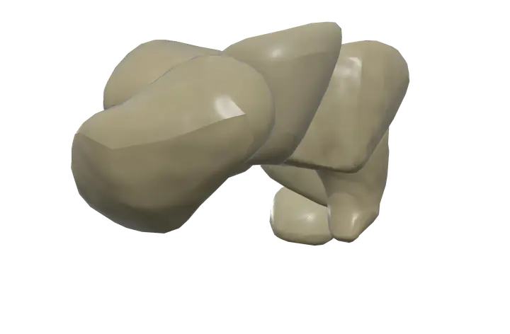
See it on the 3D body
The carpal bones lit on the 3D forearm and hand, as in the forearm and hand quiz. Turn the body, peel the layers, tap any structure for its name.
Quick answer
- Latin name
- Ossa carpi
- Type
- Short bones (eight), the pisiform also a sesamoid bone
- Location
- Wrist, between the radius and ulna and the five metacarpals
- Joints
- Wrist (radiocarpal) joint with the radius; midcarpal and intercarpal joints between the carpals; carpometacarpal joints with the metacarpals
The carpal bones are the eight small bones of the wrist, set in two rows of four between the forearm and the palm: the scaphoid, lunate, triquetrum and pisiform in the proximal row, and the trapezium, trapezoid, capitate and hamate in the distal row. Together they make the wrist both flexible and strong enough to pass force from the hand to the forearm.
Where they are
The carpals fill the space between the lower ends of the radius and ulna and the bases of the five metacarpals. They are packed closely, joined by many short ligaments, and slide a little against each other when the wrist moves.
In Anatomy Atlas 3D the eight carpals of each wrist are shown as one structure, "Carpal bones", so you see the whole block at once. The sections below describe the eight bones inside it.
The eight carpal bones
Listed in each row from the thumb side (lateral) to the little-finger side (medial):
| Row | Bone | Shape and key feature |
|---|---|---|
| Proximal | Scaphoid | Boat-shaped, the largest of the proximal row. Its tubercle can be felt at the base of the thumb. |
| Proximal | Lunate | Half-moon shaped, in the center of the row, facing the radius. |
| Proximal | Triquetrum | Pyramid-shaped, with a small facet for the pisiform. |
| Proximal | Pisiform | Pea-shaped, the smallest carpal, sitting on the front of the triquetrum. |
| Distal | Trapezium | Four-sided, with a saddle surface for the thumb metacarpal and a tubercle on its front. |
| Distal | Trapezoid | Small and wedge-shaped, behind the index metacarpal. |
| Distal | Capitate | The largest carpal, with a rounded head, in the center of the wrist. |
| Distal | Hamate | Wedge-shaped, with a curved hook projecting toward the palm. |
The pisiform develops inside the tendon of the flexor carpi ulnaris, so it is also classed as a sesamoid bone, like the patella at the knee.
Joints of the wrist
- Radiocarpal (wrist) joint. The scaphoid and lunate, with the triquetrum at the edge, meet the radius and a disc of cartilage below the ulna. The ulna itself does not touch the carpals. This joint allows the wrist to bend forward (flexion), back (extension) and side to side (radial and ulnar deviation).
- Midcarpal joint. Between the proximal and distal rows. It adds a large share of wrist flexion and extension.
- Intercarpal joints. Between neighboring bones in the same row; only small gliding movements.
- Carpometacarpal joints. Between the distal row and the metacarpals. The one between the trapezium and the first metacarpal is a saddle joint that lets the thumb swing across the palm to touch the other fingertips.
- The pisiform has only one joint, with the front of the triquetrum.
The carpal arch and the carpal tunnel
Seen from the palm, the carpals form an arch, hollow on the front, called the carpal groove. Four bony pillars stand at its corners: the pisiform and the hook of the hamate on the little-finger side, and the tubercle of the scaphoid and the tubercle of the trapezium on the thumb side.
A strong band, the flexor retinaculum, spans these pillars and closes the groove into the carpal tunnel. Through it pass the median nerve and the long flexor tendons of the fingers and thumb, on their way from the muscles of the forearm to the hand.
Muscles attached to the carpal bones
The carpals are not big muscle anchors, but several attach to them:
- The flexor carpi ulnaris inserts on the pisiform, and through ligaments continues to the hook of the hamate and the fifth metacarpal.
- The abductor digiti minimi of the hand starts on the pisiform.
- The opponens digiti minimi and flexor digiti minimi brevis start on the hook of the hamate and the flexor retinaculum.
- The short thumb muscles of the thenar eminence start on the flexor retinaculum and the tubercles of the scaphoid and trapezium.
- The oblique head of the adductor pollicis starts on the capitate and the bases of the nearby metacarpals.
Most of the long muscles that move the wrist, such as the flexor carpi radialis or the wrist extensors, cross the carpals and insert on the metacarpals instead.
How to remember them
- A mnemonic. "Sally Left The Party To Take Cathy Home": Scaphoid, Lunate, Triquetrum, Pisiform, Trapezium, Trapezoid, Capitate, Hamate. Start at the thumb side of the proximal row, then go back to the thumb side for the distal row.
- Count the pillars. Pisiform and hamate on the little-finger side, scaphoid and trapezium on the thumb side: these four hold the flexor retinaculum.
- Thumb pairs. The trapezium carries the thumb; the scaphoid sits just behind it.
The bones of the hand page puts the carpals in context with the metacarpals and phalanges.
Frequently asked questions
What are the 8 carpal bones in order?
Proximal row, thumb to little finger: scaphoid, lunate, triquetrum, pisiform. Distal row, thumb to little finger: trapezium, trapezoid, capitate, hamate.
Which carpal bone is the largest?
The capitate, in the center of the distal row. The smallest is the pisiform.
Which carpal bones form the wrist joint with the radius?
Mainly the scaphoid and the lunate, with the triquetrum touching the disc of cartilage beneath the ulna.
Which bones form the carpal tunnel?
The carpal groove formed by the eight carpals, with the pisiform, the hook of the hamate and the tubercles of the scaphoid and trapezium as its corners. The flexor retinaculum forms the roof.
Why are the carpal bones one structure in the app?
The app shows the eight bones of each wrist together as "Carpal bones", so one tap highlights the whole wrist block. The table above names the eight bones inside it.
Muscles that originate here
Muscles that insert here
From the anatomical data behind Anatomy Atlas 3D (Z-Anatomy, reviewed). In the app, tap the bone, then each muscle around it to open its card.
