
Joint · Lower limb
Knee joint anatomy: bones, ligaments, menisci and muscles
The knee joint links the femur, tibia and patella in one capsule. Learn its three articulations, four main ligaments, two menisci and the muscles that move it.
The knee joint is the largest joint of the body, where the femur meets the tibia and the patella. It works mainly as a hinge: two ligaments on its sides and two crossed ligaments inside hold it together, two menisci cushion it, the quadriceps straightens it and the hamstrings bend it.
Which bones form the knee joint?
Three bones meet at the knee:
- The femur, whose two rounded condyles rest on the tibia, with a smooth groove in front of them, the patellar surface, for the kneecap.
- The tibia, whose medial and lateral condyles form a nearly flat top. Between them rises the intercondylar eminence, where the two cruciate ligaments are anchored.
- The patella, a sesamoid bone inside the tendon of the quadriceps, which glides up and down in the femoral groove.
The fibula is not part of the knee joint. Its head sits just below the knee on the outer side and joins the tibia, not the femur, but two knee structures attach to it: the fibular collateral ligament and the biceps femoris.
What are the three articulations of the knee?
The knee is one joint with one capsule, but three pairs of surfaces move against each other inside it:
| Articulation | Between | Movement |
|---|---|---|
| Medial tibiofemoral joint | Medial condyle of the femur and medial condyle of the tibia | Rolling and gliding in flexion and extension |
| Lateral tibiofemoral joint | Lateral condyle of the femur and lateral condyle of the tibia | Rolling and gliding in flexion and extension |
| Patellofemoral joint | Back of the patella and the patellar surface of the femur | The patella slides up and down in its groove |
The patella never touches the tibia. It is linked to it by the patellar ligament, which runs from the lower tip of the patella to the tibial tuberosity, the bump at the top of the shin.
What are the ligaments of the knee?
Four ligaments do most of the work of holding the femur on the tibia:
| Ligament | Where it runs | What it does |
|---|---|---|
| Fibular (lateral) collateral ligament | Outside the capsule, from the lateral epicondyle of the femur to the head of the fibula | Taut when the knee is straight; limits side-to-side movement |
| Tibial (medial) collateral ligament | Outside the capsule, from the medial epicondyle of the femur to the medial tibia; fused to the capsule and the medial meniscus | Taut when the knee is straight; limits side-to-side movement |
| Anterior cruciate ligament (ACL) | Inside the knee, attached in front of the intercondylar eminence of the tibia, up to the inner side of a femoral condyle | Tightens in extension and resists hyperextension |
| Posterior cruciate ligament (PCL) | Inside the knee, attached behind the intercondylar eminence of the tibia, up to the inner side of the other femoral condyle | The stronger of the two; stops the femur sliding forward off the tibia when the knee is bent under load |
"Cruciate" means crossed: seen from the front, the ACL and PCL make an X in the middle of the joint. Extra ligaments woven into the back of the capsule add to the protection against hyperextension.
What do the menisci do?
A rounded femoral condyle sitting on a flat tibial condyle would touch it over a small area. The two menisci, medial and lateral, make up for that: they are wedges of fibrocartilage that deepen each tibial condyle and cushion the load. What to remember:
- Shape: each is a C, wedge-shaped in cross section, with its thick edge facing outward.
- Attachment: to the tibia only; the femur rolls on top of them.
- Mobility: the lateral meniscus moves more freely. The medial one is held at its outer edge by the capsule and the tibial collateral ligament.
- Blood supply: not all of each meniscus receives arterial blood, which matters for how well it heals.
Which muscles move the knee?
The knee bends (flexion) and straightens (extension). When it is bent, the leg can also rotate a little; when it is straight, it cannot.
| Movement | Muscles | Where they attach below the knee |
|---|---|---|
| Extension | The quadriceps femoris: rectus femoris, vastus lateralis, vastus medialis, vastus intermedius | Patella, then the tibial tuberosity through the patellar ligament |
| Flexion | The hamstrings: biceps femoris, semitendinosus, semimembranosus | Head of the fibula (biceps femoris), medial tibia (the other two) |
| Flexion, helping | Sartorius, gracilis, gastrocnemius | Medial tibia (sartorius, gracilis); the gastrocnemius crosses the back of the knee from the femur to the heel |
| Medial rotation of the bent knee | Gracilis, semimembranosus, semitendinosus | Medial tibia |
The quadriceps also steadies the knee, which is why OpenStax calls it a "dynamic ligament". The tendons of the three hamstrings frame the popliteal fossa, the diamond-shaped hollow at the back of the knee. The muscles of the thigh table lists the origin, insertion and action of the thigh muscles.
How does the knee lock when you stand?
When you stand with your knees fully straight, you can relax your thigh muscles without the knees giving way. The knee is locked. The lock comes from a small twist: the two femoral condyles are not the same size, so in the last degrees of straightening the femur rotates slightly inward on the tibia. Bending starts with the opposite twist, outward, made by the popliteus, a short muscle deep at the back of the knee.
How to study the knee joint in 3D
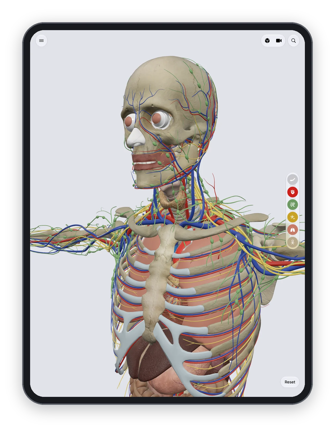
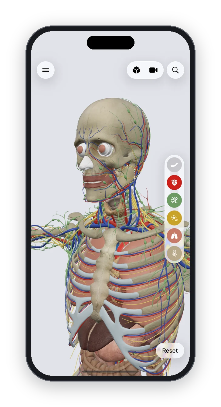
Anatomy Atlas 3D is a free 3D human anatomy atlas for iPad and iPhone: 2,000 structures, no account, no ads, works offline, English and Latin names, and a tap-the-structure quiz. It shows the bones and muscles of the knee, not its ligaments or menisci, so use it for the part of the knee that moves:
- Peel away the muscle layer and turn the skeleton to the back of the knee. Look at the two femoral condyles, the intercondylar fossa between them and the flat top of the tibia. The fossa is where the cruciate ligaments sit; the menisci rest on the flat tibial condyles.
- Turn back to the front and follow the line from the quadriceps to the patella and down to the tibial tuberosity.
- Bring the muscles back and tap each hamstring and the gastrocnemius to see where it crosses the knee.
- Test yourself with the hip and thigh quiz or the lower limb muscles quiz, and find every lower limb page on the lower limb hub.
Frequently asked questions
What type of joint is the knee?
A synovial joint that works mainly as a hinge, allowing flexion and extension, with a little rotation of the leg when the knee is bent.
What are the four main ligaments of the knee?
The anterior and posterior cruciate ligaments inside the joint, and the tibial (medial) and fibular (lateral) collateral ligaments on its sides.
Is the fibula part of the knee joint?
No. The knee joint is formed by the femur, the tibia and the patella. The head of the fibula sits below it and anchors the fibular collateral ligament and the biceps femoris.
Which muscles straighten the knee and which bend it?
The four muscles of the quadriceps femoris straighten (extend) the knee. The three hamstrings bend (flex) it, helped by the sartorius, gracilis and gastrocnemius.
What is the difference between the menisci and the cruciate ligaments?
The menisci are two C-shaped discs of fibrocartilage that pad the space between the femur and the tibia. The cruciate ligaments are two bands that cross in the middle of the joint and tie the femur to the tibia.
