
Muscle · Trunk
Internal intercostal muscles: origin, insertion, action and innervation
The internal intercostals run from the costal groove of each rib to the rib below. They lower the ribs in forced expiration; nerves: intercostal (T1–T11).
Quick answer
- Latin name
- Musculi intercostales interni
- Origin
- Floor of the costal groove and lower border of each rib, from rib 1 to rib 11
- Insertion
- Upper border of the rib below
- Action
- Interosseous (lateral) parts depress the ribs in forced expiration; interchondral parts, between the costal cartilages, help elevate the ribs
- Innervation
- Intercostal nerves (anterior rami of T1–T11)
- Blood supply
- Posterior intercostal arteries, anterior intercostal arteries (from the internal thoracic and musculophrenic arteries)
See it on the 3D body
The internal intercostal lit on the 3D thorax, as in the thorax quiz. Turn the body, peel the layers, tap any structure for its name.
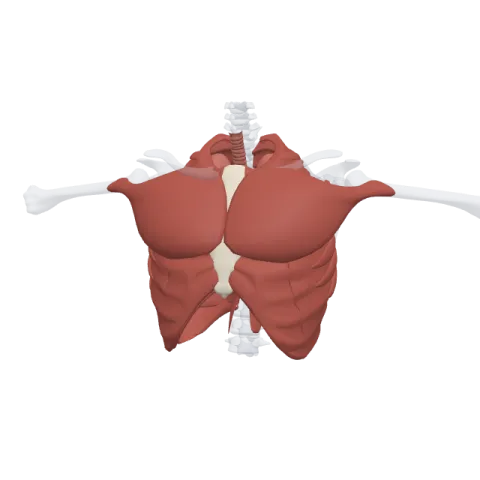
The internal intercostals are eleven pairs of thin muscles that form the middle layer of each space between the ribs, just deep to the external intercostals. Their interosseous parts, between the bony ribs, pull the ribs down when you breathe out hard. Each one gets its nerve from the intercostal nerve running in the same space.
Where it is
Each intercostal space holds three layers of muscle. From outside in: the external intercostals, the internal intercostals and a thin, incomplete innermost layer. The internal intercostals sit in the middle, covered by the external layer on the outside and lined on the inside by the innermost intercostals and the pleura.
They cover the opposite stretch of the chest from the external layer. The internal intercostals start at the front, right next to the sternum, and run back around the chest as far as the angle of the rib. Behind that point, toward the spine, the fibers give way to a thin sheet, the internal intercostal membrane. So in the front of the chest, between the costal cartilages, the internal intercostal is the only one of the three layers that still has muscle fibers.
The fibers run down and back, at right angles to the external intercostals. On each side of the chest the two layers cross like an X.
Origin and insertion
- Origin: ribs 1 to 11, along the floor of the costal groove and the lower edge of each rib.
- Insertion: the upper border of the rib directly below.
Anatomists split each muscle into two parts by position:
- The interosseous part, between the bony parts of two ribs, on the side and front of the chest.
- The interchondral part, between two costal cartilages, close to the sternum.
What it does
The interosseous and interchondral parts do different jobs, and the whole layer also keeps the spaces firm:
- Interosseous parts: forced expiration. They pull each rib down toward the rib below. This narrows the chest and pushes air out. You use them when you blow out candles, cough or play a wind instrument. In quiet breathing, breathing out is mostly passive: the lungs and chest wall recoil on their own.
- Interchondral parts: help with inspiration. Between the cartilages near the sternum, the fibers act like the external intercostals and help lift the ribs.
- Keeping the spaces firm. Like the outer layer, they stop the soft tissue between the ribs from bulging or caving in as the pressure in the chest changes.
Innervation and blood supply
Each internal intercostal is supplied by the intercostal nerve of its space, from the anterior rami of T1 to T11. The intercostal vein, artery and nerve run together just under each rib, in the costal groove, in the plane between the internal and innermost intercostals. From top to bottom they lie in the order vein, artery, nerve.
The muscles get their blood from the posterior intercostal arteries and the anterior intercostal arteries, which meet in each space.
How to remember it
- Internal = in from the front. The internal layer starts beside the sternum and stops at the rib angle; the external layer starts at the back and stops at the cartilage.
- Crossed arms: external fibers point down and forward, internal fibers down and back. Cross your hands on your chest and they form the same X.
- VAN under the rib: Vein, Artery, Nerve, from top to bottom, tucked into the costal groove between the internal and innermost layers.
- Forced out, not quiet out: the interosseous parts work when you blow hard, not when you breathe calmly.
Practice spotting both layers and the ribs in the thorax quiz, and review the ribs themselves on the bones of the rib cage page.
Frequently asked questions
What do the internal intercostal muscles do?
Their interosseous parts pull the ribs down during forced expiration, such as coughing or blowing. Their interchondral parts, near the sternum, help lift the ribs.
Are the internal intercostals used in normal breathing?
Not much. Quiet breathing out is mostly passive recoil of the lungs and chest wall. The internal intercostals join in when you breathe out with force.
Which way do the internal intercostal fibers run?
Down and back, at right angles to the external intercostal fibers.
Where is the intercostal neurovascular bundle?
In the costal groove under each rib, between the internal intercostal and the innermost intercostal layer, in the order vein, artery, nerve from top to bottom.
