
Muscle · Trunk
External intercostal muscles: origin, insertion, action and innervation
The external intercostals run from each rib to the rib below it. They lift the ribs to breathe in and are supplied by the intercostal nerves (T1–T11).
Quick answer
- Latin name
- Musculi intercostales externi
- Origin
- Lower border of each rib, from rib 1 to rib 11
- Insertion
- Upper border of the rib below
- Action
- Elevate the ribs during inspiration; stiffen the intercostal spaces
- Innervation
- Intercostal nerves (anterior rami of T1–T11)
- Blood supply
- Posterior intercostal arteries, anterior intercostal arteries (from the internal thoracic and musculophrenic arteries)
See it on the 3D body
The external intercostal lit on the 3D thorax, as in the thorax quiz. Turn the body, peel the layers, tap any structure for its name.
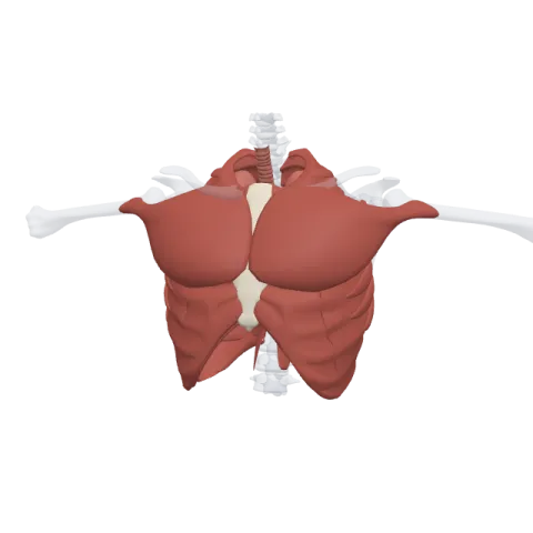
The external intercostals are eleven pairs of thin muscles that fill the outer layer of each space between two ribs. They pull each rib up toward the one above, which widens the chest when you breathe in, and they are supplied by the intercostal nerves.
Where it is
Between any two neighboring ribs there are three thin sheets of muscle, stacked like plywood. The external intercostals are the outermost sheet, just under the chest muscles such as the pectoralis major and the serratus anterior.
Each muscle starts at the back, near the tubercle of the rib, and runs forward around the chest. It stops at about the point where the bony rib meets its costal cartilage. From there to the sternum, the muscle fibers are replaced by a thin sheet of connective tissue, the external intercostal membrane. So at the front of the chest, close to the breastbone, you find no external intercostal fibers at all.
The fibers run down and forward, in the same direction as the external oblique of the abdomen. If you put your hands in your front pockets, your fingers point the way the external intercostal fibers run.
Origin and insertion
- Origin: the lower border of a rib, from the first to the eleventh.
- Insertion: the upper border of the rib directly below.
Because the thorax has twelve pairs of ribs, it has eleven intercostal spaces on each side, and so eleven external intercostal muscles per side. Below the twelfth rib there is no space to fill: the abdominal muscles take over.
What it does
The external intercostals are muscles of inspiration. When they contract, each rib is pulled up toward the rib above. Because the ribs slope downward from the spine, lifting them pushes the sternum forward (the "pump handle" movement of the upper ribs) and swings the lower ribs outward (the "bucket handle" movement). Both enlarge the chest from front to back and from side to side, so air flows into the lungs.
In quiet breathing, the diaphragm does most of the work and the external intercostals help. When you breathe in deeply, as after a run, they work harder. They also keep the intercostal spaces firm, so the soft tissue between the ribs is not sucked in or pushed out as the pressure inside the chest changes.
Innervation and blood supply
Each external intercostal muscle is supplied by the intercostal nerve of its space. The intercostal nerves are the anterior rami of the thoracic spinal nerves T1 to T11. The nerve, the posterior intercostal artery and the intercostal vein travel together in the costal groove, under the lower edge of each rib, between the inner two layers of muscle.
The blood comes from the posterior intercostal arteries, which arise from the thoracic aorta (the first two come from a branch of the subclavian artery), and from the anterior intercostal arteries, branches of the internal thoracic artery and its musculophrenic branch.
How to remember it
- External = expand. Both start with "ex": the external intercostals expand the chest to breathe in.
- Hands in pockets: the fibers point down and forward, like the external oblique, which continues the same outer layer on the abdomen.
- Stops at the cartilage: external fibers fade out near the costal cartilages and become the external intercostal membrane in front.
- Count the gaps: twelve ribs, eleven spaces, eleven external intercostals per side.
Test yourself on the chest wall, ribs and lungs in the thorax quiz, and see how the ribs are built on the bones of the rib cage page.
Frequently asked questions
What is the main function of the external intercostal muscles?
They lift the ribs during inspiration, which enlarges the chest and draws air into the lungs. They help the diaphragm, especially when you breathe in deeply.
Which way do the external intercostal fibers run?
Down and forward, from the rib above to the rib below. The internal intercostals run at right angles to them.
What nerve supplies the external intercostals?
The intercostal nerves, which are the anterior rami of T1 to T11. Each muscle is supplied by the nerve of its own space.
How many external intercostal muscles are there?
Eleven on each side, one in each intercostal space, so twenty-two in total.
What is the difference between the external and internal intercostals?
The external layer is outside, runs down and forward and lifts the ribs to breathe in. The internal layer is just inside it, runs down and back, and its side parts lower the ribs during forced breathing out.
