
Muscle · Trunk
Internal oblique muscle: origin, insertion, action and innervation
The internal oblique runs from the iliac crest, inguinal ligament and lumbar fascia to ribs 10–12 and the linea alba. It turns the trunk; T7–L1 nerves.
Quick answer
- Latin name
- Musculus obliquus internus abdominis
- Origin
- Thoracolumbar fascia, anterior two thirds of the iliac crest of the hip bone, lateral part of the inguinal ligament
- Insertion
- Lower borders of ribs 10–12, linea alba (through its aponeurosis), pubic crest and pecten pubis (through the conjoint tendon)
- Action
- Compresses the abdominal viscera; flexes the trunk; bends it to the same side; rotates it to the same side, with the opposite external oblique
- Innervation
- Thoraco-abdominal nerves (T7–T11), subcostal nerve (T12), iliohypogastric and ilioinguinal nerves (L1)
- Blood supply
- Lower posterior intercostal and subcostal arteries, superior and inferior epigastric arteries, deep circumflex iliac artery
See it on the 3D body
The internal abdominal oblique lit on the 3D abdomen, as in the abdomen quiz. Turn the body, peel the layers, tap any structure for its name.
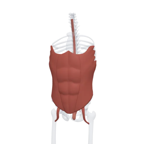
The internal oblique is the middle of the three flat muscles on the side of the abdomen, lying between the external oblique and the transversus abdominis. It squeezes the abdominal contents and turns the trunk toward its own side, and it is supplied by the lower thoracic nerves and the first lumbar nerve.
Where it is
Picture the side of the abdomen as three sheets of muscle laid one on top of the other. The external oblique is outside. The internal oblique is in the middle. The transversus abdominis is the deepest. All three turn into flat tendons (aponeuroses) toward the front, and these wrap around the rectus abdominis to form the rectus sheath before meeting at the midline in the linea alba.
The fibers of the internal oblique fan out from the hip and lower back. Most run up and forward, at right angles to the external oblique. The lowest fibers run almost horizontally and then curve down toward the pubis. In males, some of these lowest fibers follow the spermatic cord into the scrotum as the cremaster muscle.
In the upper part of the abdomen, the aponeurosis of the internal oblique splits in two around the rectus abdominis: one sheet passes in front of the rectus, the other behind it. Below a curved line called the arcuate line, all of it passes in front.
Origin and insertion
- Origin: the thoracolumbar fascia in the lower back, the front two thirds of the iliac crest of the hip bone, and the lateral part of the inguinal ligament.
- Insertion: the lower borders of ribs 10 to 12, the linea alba through its aponeurosis, and the pubic crest and pecten pubis through the conjoint tendon, which it shares with the transversus abdominis.
What it does
- Compresses the abdomen. With the other flat muscles, it raises the pressure inside the abdomen. This helps you breathe out hard, cough, sneeze, vomit, push during childbirth and empty the bladder and bowel.
- Flexes the trunk when the muscles of both sides contract, working with the rectus abdominis.
- Bends the trunk to the same side when one side contracts.
- Rotates the trunk to the same side. The left internal oblique turns the chest to the left. It does this together with the right external oblique, whose fibers run in the same line on the other side of the body.
Innervation and blood supply
The internal oblique is supplied by the thoraco-abdominal nerves (the lower intercostal nerves, T7 to T11), the subcostal nerve (T12), and the iliohypogastric and ilioinguinal nerves (L1). These nerves run forward in the plane between the internal oblique and the transversus abdominis, which is why that plane is called the neurovascular plane of the abdominal wall.
Its blood comes from the lower posterior intercostal and subcostal arteries, the superior and inferior epigastric arteries and the deep circumflex iliac artery.
How to remember it
- Same-side turner. Internal oblique turns you toward the internal side, your own side: left internal, turn left.
- Diagonal partners. The right external oblique and the left internal oblique form one long diagonal line across the front of the trunk, so they work as a team when you twist.
- External in pockets, internal up the ribs. If external oblique fibers are hands sliding into front pockets, internal oblique fibers point the other way, up toward the lower ribs.
- Ribs 10 to 12 on top. Of the three flat muscles, it is the one whose upper fibers end on the lower borders of the last three ribs.
Compare all four abdominal wall muscles on the abdominal muscles page, then test yourself in the abdomen quiz.
Frequently asked questions
What is the action of the internal oblique?
It compresses the abdominal contents, flexes the trunk, bends it to the same side and rotates it to the same side, working with the external oblique of the opposite side.
Which way do the internal oblique fibers run?
Mostly up and forward, at right angles to the external oblique. The lowest fibers run horizontally and then curve down toward the pubis.
What nerves supply the internal oblique?
The thoraco-abdominal nerves (T7 to T11), the subcostal nerve (T12) and the iliohypogastric and ilioinguinal nerves (L1).
What is the conjoint tendon?
The combined lower ends of the internal oblique and transversus abdominis aponeuroses, which attach to the pubic crest and pecten pubis behind the inguinal canal.
Is the cremaster muscle part of the internal oblique?
It comes from it. The cremaster is formed by the lowest fibers of the internal oblique, which follow the spermatic cord down to the testis.
