
Muscle · Trunk
Diaphragm: origin, insertion, action and innervation
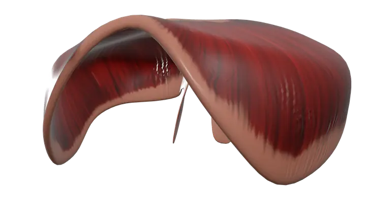
The diaphragm is the main breathing muscle, from the xiphoid, lower six ribs and L1-L3 to its central tendon. It is supplied by the phrenic nerves.
Quick answer
- Latin name
- Diaphragma
- Origin
- Sternal part: back of the xiphoid process of the sternum. Costal part: inner surfaces of the lower six costal cartilages and ribs. Lumbar part: crura on the bodies of L1–L3 and the arcuate ligaments
- Insertion
- Central tendon
- Action
- Main muscle of inspiration: contracts and flattens, enlarging the chest cavity; helps raise pressure inside the abdomen
- Innervation
- Phrenic nerves (C3–C5); the lower intercostal nerves carry sensation from its outer edge
- Blood supply
- Superior and inferior phrenic arteries, musculophrenic and pericardiacophrenic arteries, and the lower intercostal arteries
See it on the 3D body
The diaphragm lit on the 3D thorax, as in the thorax quiz. Turn the body, peel the layers, tap any structure for its name.
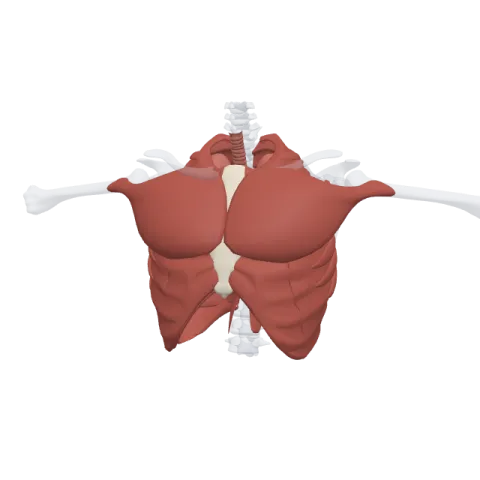
The diaphragm is the dome-shaped sheet of muscle and tendon that separates the chest (thoracic cavity) from the abdomen. It is the main muscle of breathing: when it contracts it flattens and pulls air into the lungs, and it is supplied by the phrenic nerves (C3 to C5).
Where it is
The diaphragm forms the floor of the thoracic cavity and the roof of the abdominal cavity. Seen from the front, it rises as two domes, one on each side, under the lungs. The right dome sits higher than the left because the liver lies under it. Between the domes, a slight dip holds the heart, whose fibrous sac (the pericardium) is fused to the top of the diaphragm.
The muscle fibers are arranged around the edge, and they all run inward and upward to a flat sheet of tendon in the middle, the central tendon. Unlike most muscles, the diaphragm has no bony insertion: it pulls on its own tendon.
Several structures pass between the chest and the abdomen through three main openings:
- Caval opening (level of T8): the inferior vena cava, through the central tendon.
- Esophageal hiatus (T10): the esophagus and the vagal trunks, through the muscle of the right crus.
- Aortic hiatus (T12): the aorta, the thoracic duct and sometimes the azygos vein, behind the diaphragm, between the two crura.
Origin and insertion
The diaphragm has a ring-shaped origin with three parts:
- Sternal part: two small slips from the back of the xiphoid process of the sternum.
- Costal part: the inner surfaces of the lower six costal cartilages and ribs (ribs 7 to 12), interlocking with the transversus abdominis. See the bones of the rib cage.
- Lumbar part: two muscular columns called crura, the right crus from the bodies of L1 to L3 and the smaller left crus from L1 and L2, plus the medial and lateral arcuate ligaments that arch over the psoas major and the quadratus lumborum.
All the fibers insert on the central tendon.
What it does
- Inspiration: when the diaphragm contracts, its domes move down and flatten. This increases the height of the thoracic cavity, lowers the pressure in the lungs and draws air in. In quiet breathing, it does most of the work, helped by the external intercostal muscles that lift the ribs.
- Expiration: in quiet breathing, breathing out is passive. The diaphragm relaxes, the stretched lungs and chest wall recoil and the domes rise back up.
- Raising abdominal pressure: when the diaphragm contracts together with the abdominal muscles while the airway is closed, the pressure in the abdomen rises. This helps in coughing, sneezing, vomiting, emptying the bladder and bowels, giving birth and lifting heavy loads.
- Helping venous return: each descent of the diaphragm lowers the pressure in the chest and raises it in the abdomen, which helps push blood back toward the heart through the inferior vena cava.
Innervation and blood supply
The diaphragm receives its motor supply from the two phrenic nerves (roots C3, C4 and C5), one for each half. They start in the neck, descend through the chest on either side of the heart and reach the diaphragm from above. This high origin is explained by development: the muscle forms in the neck region of the embryo and descends later, bringing its nerve with it. The phrenic nerves also carry sensation from the central part; the outer edge receives sensory fibers from the lower intercostal nerves.
Its blood supply comes from above and below: the superior phrenic arteries (from the thoracic aorta), the inferior phrenic arteries (from the abdominal aorta), the musculophrenic and pericardiacophrenic arteries (from the internal thoracic artery) and the lower intercostal arteries.
How to remember it
- "C3, 4, 5 keeps the diaphragm alive": the roots of the phrenic nerve.
- "I 8 10 Eggs At 12": Inferior vena cava at T8, Esophagus at T10, Aorta at T12.
- Right dome higher: the liver pushes it up.
- Contract = flatten = breathe in; relax = dome rises = breathe out.
The abdominal muscles page shows how the diaphragm forms the top of the abdominal wall.
Frequently asked questions
What is the main function of the diaphragm?
It is the main muscle of inspiration. When it contracts, it flattens and enlarges the chest cavity, which draws air into the lungs.
Which nerve supplies the diaphragm?
The phrenic nerves, from roots C3, C4 and C5, one on each side.
What passes through the diaphragm?
Mainly the inferior vena cava (at T8), the esophagus with the vagal trunks (at T10) and the aorta with the thoracic duct (at T12).
Is the diaphragm a skeletal muscle?
Yes. It is a skeletal muscle, so it can be controlled voluntarily, as when you hold your breath, but it also works automatically under the control of the brainstem.
Why is the right side of the diaphragm higher?
Because the liver lies directly under the right dome and pushes it upward.
