
See it on the 3D body
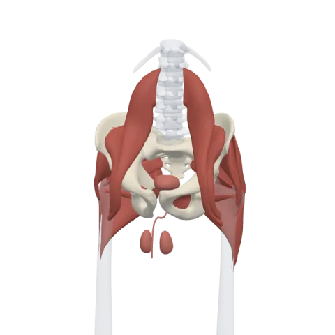
The testis lit on the 3D pelvis, as in the pelvis quiz. Turn the body, peel the layers, tap any structure for its name.
Quick answer
- Latin name
- Testis
- System
- Male reproductive system and endocrine system
- Location
- Scrotum, outside the abdominal cavity, below the root of the penis; each testis hangs from a spermatic cord
- Function
- Produces sperm in the seminiferous tubules and the hormone testosterone in the interstitial (Leydig) cells
The testes (singular: testis) are two oval glands that hang in the scrotum, outside the abdominal cavity. Each one has two jobs: it produces sperm and it produces testosterone, the main male sex hormone. So the testis belongs both to the reproductive system and to the endocrine system.
Where it is
The two testes sit in the scrotum, a pouch of skin and thin muscle below the front of the pelvis. Each adult testis is about 4 to 5 cm long.
Each testis hangs from a spermatic cord, a bundle that runs up through the inguinal canal in the lower abdominal wall, just above the pubic part of the hip bone. The cord carries the ductus deferens, the testicular artery and veins, lymph vessels and nerves, wrapped in thin layers of tissue and muscle.
Being outside the abdomen keeps the testes a few degrees cooler than the core of the body, which sperm production needs. Two muscles help adjust their temperature. The cremaster muscle in the spermatic cord pulls the testis up toward the body when it is cold. The dartos muscle in the wall of the scrotum wrinkles the skin to reduce heat loss.
Structure
Coverings
From the outside in, each testis is wrapped in:
- the tunica vaginalis, a thin, two-layered serous sac that lets the testis slide inside the scrotum;
- the tunica albuginea, a tough white capsule of dense connective tissue.
The tunica albuginea sends partitions inward that divide the testis into several hundred small lobules.
Seminiferous tubules
Each lobule contains one to a few tightly coiled seminiferous tubules, where sperm are made. Their walls hold two kinds of cells:
- germ cells, which divide and mature into sperm;
- Sertoli cells, tall support cells that feed the developing sperm and form a barrier between the blood and the inside of the tubule.
Interstitial cells
Between the tubules lie small clusters of Leydig cells, also called interstitial cells. They produce testosterone and release it into the blood.
The duct system
The tubules drain into a network of channels at the back of the testis, the rete testis. From there, small efferent ducts lead into the epididymis, a long, tightly coiled tube lying along the back of the testis, with a head at the top, a body and a tail at the bottom. Sperm finish maturing and are stored in the epididymis. From its tail, the ductus deferens carries them up through the spermatic cord into the pelvis, toward the prostate.
Vessels and nerves
The testes start to form high in the abdomen, near the kidneys, and move down into the scrotum before birth. Their vessels keep this origin:
- The testicular arteries come straight off the abdominal aorta, just below the renal arteries.
- The veins form a network around the artery in the cord, the pampiniform plexus, which cools the incoming blood. The right testicular vein drains into the inferior vena cava, the left one into the left renal vein, near the kidneys.
- Lymph from the testis drains to lymph nodes beside the aorta, not to the groin.
What it does
- Produces sperm. Sperm production, or spermatogenesis, starts at puberty and goes on throughout adult life in the seminiferous tubules.
- Produces testosterone. Testosterone drives the development of the male reproductive organs before birth and the changes of puberty, and it maintains sperm production in adults.
- Answers to the brain. The pituitary gland releases two hormones: LH, which tells the Leydig cells to make testosterone, and FSH, which acts on the Sertoli cells to support sperm production. The pituitary is described on the endocrine glands page.
How to study it
- Two products, two places. Sperm are made inside the tubules; testosterone is made between them.
- Inside out. Tubules, rete testis, efferent ducts, epididymis, ductus deferens: follow the path a sperm cell takes.
- Remember the descent. The testis began near the kidney, so its artery comes from the aorta and the left vein drains into the left renal vein.
In Anatomy Atlas 3D, look at the pelvis from the front and tap each testis to read its name, then turn the body to see how far it sits below the abdomen. Test yourself in the pelvis quiz. The app is free on iPad and iPhone.
Frequently asked questions
What does the testis produce?
Sperm, in the seminiferous tubules, and the hormone testosterone, in the Leydig cells between the tubules.
Why are the testes outside the body?
Sperm production works best a few degrees below core body temperature, and the scrotum keeps the testes cooler.
Where does the testicular artery come from?
From the abdominal aorta, just below the renal arteries, because the testis develops in the abdomen before descending.
