Quick answer
- System
- Urinary system
- Location
- Kidneys high on the back wall of the abdomen, bladder in the pelvis behind the pubic bones
- Function
- Filter the blood, make urine and store it until it is released
- Parts in the app
- 7
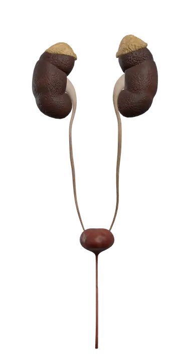
The urinary system cleans the blood. The two kidneys filter it and turn waste and extra water into urine, the two ureters carry the urine down, and the bladder stores it until it leaves the body through the urethra. The app also shows the two adrenal glands, small hormone glands that sit on top of the kidneys.
Where it is
The kidneys lie against the back wall of the abdomen, one on each side of the spine, behind the peritoneum (they are retroperitoneal). They sit roughly between the last thoracic and the third lumbar vertebrae, and their upper ends are partly protected by the lowest ribs. The right kidney sits a little lower than the left, because the liver takes up space above it. Each kidney is wrapped in a cushion of fat.
On the inner, concave side of each kidney is the hilum, where the renal artery enters and the renal vein and the ureter leave.
The ureters run down the back wall of the abdomen, cross into the pelvis and enter the back of the bladder at an angle.
The bladder sits in the pelvis, just behind the pubic symphysis, where the two hip bones meet at the front. When it fills, it rises upward into the abdomen.
The adrenal glands cap the upper end of each kidney. The right one is roughly triangular, the left one more crescent-shaped.
Parts you can explore in the app
In the app you can open these organs on their own and study them part by part:
- Left kidney: a bean-shaped organ that filters blood and makes urine.
- Right kidney: the same, sitting slightly lower than the left.
- Left ureter: a thin muscular tube that carries urine from the left kidney to the bladder.
- Right ureter: the same tube on the right side.
- Bladder: a hollow, stretchy muscular bag in the pelvis that stores urine.
- Left adrenal gland: a small endocrine gland on top of the left kidney.
- Right adrenal gland: a small endocrine gland on top of the right kidney.
Cross sections (sagittal, coronal and axial) let you cut through a kidney and see its outer cortex, its inner medulla and the cavity where urine collects.
What it does
The kidneys each contain a very large number of tiny filtering units called nephrons. In each nephron, a knot of capillaries, the glomerulus, filters fluid out of the blood. As this fluid flows along the renal tubule, water, glucose, salts and other useful substances are taken back into the blood, and some waste is added. What remains is urine.
Urine collects in small cups called calyces, drains into the funnel-shaped renal pelvis and leaves through the ureter.
By adjusting what they keep and what they remove, the kidneys also:
- control the volume of blood and help regulate blood pressure, partly through the enzyme renin;
- keep levels of sodium, potassium and other ions stable;
- help balance the acidity of the blood;
- release erythropoietin, which stimulates red blood cell production;
- convert vitamin D into its active form.
The ureters move urine by waves of muscle contraction (peristalsis), not just by gravity. Because they pass through the bladder wall at a slant, the wall presses them shut as the bladder fills, which helps stop urine from flowing back up.
The bladder has a thick muscular wall, the detrusor muscle, and an inner lining that unfolds as it fills. A smooth triangle on its floor, the trigone, is marked by the two ureter openings and the exit to the urethra. An internal sphincter works automatically, while an external sphincter lets you control when you urinate.
The adrenal glands belong to the endocrine system. Their outer cortex makes aldosterone (salt and water balance), cortisol (stress response and blood glucose) and small amounts of androgens. Their inner medulla releases epinephrine and norepinephrine in fight-or-flight situations.
How to study it
- Ureter versus urethra. There are two ureters (kidney to bladder) and one urethra (bladder to outside). Mixing them up is the most common slip.
- Trace a drop of urine. Nephron, collecting duct, minor calyx, major calyx, renal pelvis, ureter, bladder, urethra.
- What passes through the hilum. Renal artery, renal vein and ureter (plus nerves and lymphatics).
- Adrenal cortex from outside in: "salt, sugar, sex." The outer zone makes aldosterone, the middle zone cortisol, the inner zone androgens.
- Remember why the right kidney is lower. The liver pushes it down.
- Use the coronal section through a kidney to see cortex, renal pyramids of the medulla and the renal pelvis in one view.
Frequently asked questions
Why is the right kidney lower than the left?
Because the liver sits above it on the right side of the abdomen and takes up space.
What is the difference between the ureters and the urethra?
The two ureters carry urine from the kidneys to the bladder. The single urethra carries urine from the bladder out of the body.
What is the trigone of the bladder?
A smooth triangular area on the floor of the bladder, with the two ureter openings and the opening of the urethra at its corners.
Are the adrenal glands part of the urinary system?
No. They sit on top of the kidneys but belong to the endocrine system, releasing hormones such as cortisol, aldosterone and epinephrine.