Quick answer
- Latin name
- Auris
- System
- Nervous system (special senses)
- Location
- Side of the head, mostly inside the temporal bone
- Function
- Turns sound into nerve signals and senses head position and movement
- Parts in the app
- 11
The ear does two jobs at once: it lets you hear, and it tells the brain where your head is and how it is moving, so you keep your balance. Most of it is hidden inside the temporal bone of the skull, which makes it hard to picture from a flat drawing.
Where it is
The ear has three regions:
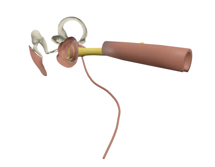
- The outer ear: the auricle (the visible flap) and the external acoustic meatus, the canal that leads inward to the eardrum.
- The middle ear: a small air-filled cavity in the temporal bone, between the eardrum and the inner ear. It holds the three ossicles and connects to the throat through the Eustachian tube.
- The inner ear: a set of fluid-filled channels carved into the dense petrous part of the temporal bone, containing the cochlea, the vestibule and the semicircular canals.
Just behind the ear you can feel the mastoid process of the temporal bone, where the sternocleidomastoid attaches.
Parts you can explore in the app
In the app you can open the ear on its own and study it part by part:
- Cochlea: the snail-shaped, coiled tube of the inner ear. Hair cells inside it turn sound vibrations into nerve signals.
- Vestibule and canals: the vestibule senses head tilt and straight-line movement; the three semicircular canals sense turning of the head.
- Malleus: the "hammer," the first ossicle, attached to the inner side of the eardrum.
- Incus: the "anvil," the middle ossicle, linking the malleus to the stapes.
- Stapes: the "stirrup," the last ossicle and the smallest bone in the body. Its footplate fits into the oval window of the inner ear.
- Eardrum: the tympanic membrane, a thin membrane between the outer and middle ear that vibrates when sound hits it.
- Eustachian tube: the auditory tube linking the middle ear to the back of the nose and throat, so that air pressure stays equal on both sides of the eardrum.
- Cochlear nerve: carries hearing signals from the cochlea.
- Vestibular nerve: carries balance signals from the vestibule and canals.
- Vestibulocochlear nerve (VIII): the eighth cranial nerve, formed by the cochlear and vestibular nerves, which runs to the brainstem.
- Chorda tympani: a branch of the facial nerve (VII) that crosses the middle ear. It carries taste from the front two-thirds of the tongue and nerve fibers for two salivary glands; it has nothing to do with hearing.
Cross sections (sagittal, coronal and axial) let you see how the canal, the middle ear and the inner ear line up inside the bone.
What it does
Hearing follows a chain of steps:
- Sound waves travel down the ear canal and make the eardrum vibrate.
- The malleus, incus and stapes pass the vibration across the middle ear. Because the eardrum is much larger than the oval window, the ossicles concentrate the force and amplify the signal.
- The stapes pushes on the oval window and sets the fluid inside the cochlea moving. The round window bulges outward to let the pressure escape.
- Waves in the fluid move the basilar membrane, bending the hair cells of the spiral organ. Different pitches stimulate different places: high sounds near the base of the cochlea, low sounds near its tip.
- The cochlear nerve carries the signals to the brainstem, and from there they travel through the thalamus to the auditory cortex of the temporal lobe.
Balance depends on the vestibular apparatus. In the vestibule, two small sacs, the utricle and the saccule, contain hair cells topped with tiny crystals that shift with gravity and straight-line acceleration. The three semicircular canals sit at roughly right angles to one another; when the head turns, the fluid inside lags behind and bends hair cells at the base of each canal. The brain combines these signals with information from the eyes and from muscles and joints.
The Eustachian tube opens briefly when you swallow or yawn, which is why swallowing helps when your ears feel blocked during a flight.
How to study it
- Three regions, three jobs. The outer ear collects sound, the middle ear amplifies it, and the inner ear converts it into nerve signals and senses balance.
- Ossicles in order: "MIS." Malleus, Incus, Stapes, from the eardrum inward.
- Two windows. The stapes pushes on the oval window; the round window releases the pressure.
- Who senses what. Cochlea = hearing; utricle and saccule = gravity and straight-line movement; semicircular canals = rotation.
- Don't mix up VII and VIII. The vestibulocochlear nerve (VIII) carries hearing and balance; the chorda tympani belongs to the facial nerve (VII) and carries taste. Nerves VII and VIII enter the internal acoustic meatus together, which makes them a frequent exam trap.
- Use the coronal section to see the canal, eardrum, ossicles and inner ear in one line.
Frequently asked questions
What are the three bones of the middle ear?
The malleus, the incus and the stapes, together called the ossicles. They carry vibrations from the eardrum to the inner ear.
What is the smallest bone in the body?
The stapes, the innermost of the three ossicles.
What does the Eustachian tube do?
It connects the middle ear to the upper throat and keeps the air pressure in the middle ear equal to the pressure outside.
How does the ear help with balance?
The vestibule senses head tilt and straight-line movement, and the semicircular canals sense rotation. The vestibular nerve sends this information to the brain.