Quick answer
- Latin name
- Encephalon
- System
- Central nervous system
- Location
- Inside the skull, filling the cranial cavity
- Function
- Processes sensation, controls movement, and supports thought, memory and emotion
- Parts in the app
- 6
The brain is the control center of the nervous system. Together with the spinal cord it forms the central nervous system, which receives information from the senses, decides what to do with it, and sends commands to muscles and glands. Thought, memory, language and emotion all depend on it.
Where it is
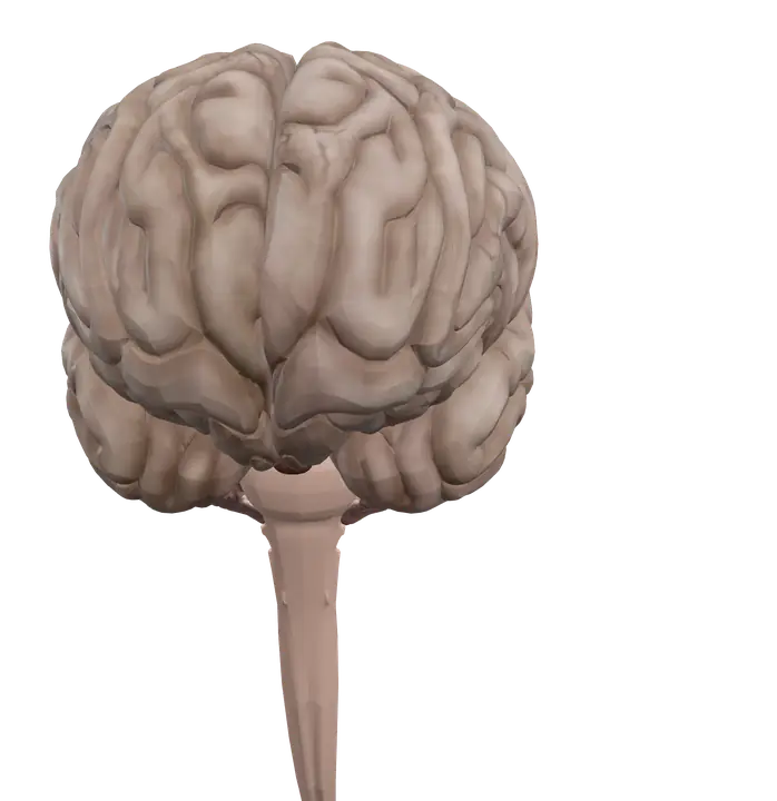
The brain fills the cranial cavity of the skull. The large cerebrum sits on top and makes up most of its volume. The cerebellum tucks in at the back, below the occipital part of the cerebrum. The brainstem runs down the middle and continues through the large opening at the base of the skull, the foramen magnum, as the spinal cord.
Three membranes, the meninges, wrap the brain: the tough dura mater on the outside, the delicate arachnoid mater in the middle and the thin pia mater stuck to the brain surface. Cerebrospinal fluid fills the space under the arachnoid and the cavities inside the brain, the brain ventricles, and cushions the brain against knocks.
The pituitary gland hangs from the underside of the brain by a thin stalk and sits in a small hollow of the sphenoid bone called the sella turcica.
Parts you can explore in the app
In the app you can open the brain on its own and study it part by part:
- Left hemisphere: the left half of the cerebrum. It receives sensation from the right side of the body and controls its movements, and in most people it carries much of the work of language.
- Right hemisphere: the right half of the cerebrum. It receives sensation from the left side of the body and controls its movements.
- Cerebellar hemisphere (left): the left half of the cerebellum, which coordinates movements of the left side of the body.
- Cerebellar hemisphere (right): the right half of the cerebellum, which coordinates movements of the right side of the body.
- Brainstem and spinal cord: the midbrain, pons and medulla oblongata, continuing into the spinal cord. This is the main highway between the brain and the body, and the site of centers that keep you breathing.
- Pituitary gland: a pea-sized gland under the brain that releases hormones controlling many other endocrine glands.
Sagittal, coronal and axial cross sections let you see the inside of the brain, including the gray matter of the surface and the white matter beneath it.
What it does
The surface of each hemisphere is the cerebral cortex, a folded sheet of gray matter. The ridges are gyri, the grooves are sulci, and deep grooves split each hemisphere into lobes:
- Frontal lobe: planning, decision-making and voluntary movement. The primary motor cortex lies in the precentral gyrus, just in front of the central sulcus. Broca's area, involved in producing speech, is usually in the left frontal lobe.
- Parietal lobe: touch, temperature, pain and body position. The primary somatosensory cortex lies in the postcentral gyrus, just behind the central sulcus.
- Temporal lobe: hearing, memory and understanding language. Wernicke's area is usually in the left temporal lobe.
- Occipital lobe: vision. Signals from the eye end up here.
Under the cortex, white matter carries signals between regions. The largest bundle, the corpus callosum, links the two hemispheres so they can share information.
Most sensory and motor pathways cross the midline on their way, which is why each hemisphere deals with the opposite side of the body. The cerebellum is different: it works with the same side. It compares the movement you intended with the movement you actually made and corrects it, keeping you balanced and smooth.
The brainstem handles vital automatic functions such as breathing, heart rate and blood pressure, as well as reflexes like swallowing and coughing. Most cranial nerves attach to it, including the nerves for the ear. The pituitary links the nervous system to the hormone system through its connection with the hypothalamus.
How to study it
- Start with three landmarks. The longitudinal fissure separates the hemispheres, the central sulcus separates frontal from parietal lobes, and the lateral sulcus separates the temporal lobe from the lobes above it.
- "Motor in front, sensory behind." The precentral gyrus is motor; the postcentral gyrus is somatosensory.
- Opposite cerebrum, same-side cerebellum. A classic exam point that also explains many clinical cases you will meet later.
- Meninges from the inside out: PAD. Pia, Arachnoid, Dura.
- Cranial nerves. Nerves III to XII attach to the brainstem; nerves I and II connect to the forebrain.
- Use the sagittal section. One midline slice shows the corpus callosum, the brainstem, the cerebellum and the pituitary together, which is the view most exams ask you to label.
Frequently asked questions
What are the main parts of the brain?
The cerebrum (two hemispheres), the diencephalon (thalamus and hypothalamus), the cerebellum and the brainstem. The brainstem continues as the spinal cord.
Why does the left side of the brain control the right side of the body?
Most motor and sensory pathways cross to the opposite side as they travel between the brain and the spinal cord, so each hemisphere handles the opposite half of the body.
What does the cerebellum do?
It coordinates movement, balance and posture, and helps with learning motor skills. It fine-tunes movements rather than starting them.
Is the pituitary gland part of the brain?
It is attached to the hypothalamus at the base of the brain. Its posterior part is an extension of nervous tissue, while its anterior part is glandular tissue.