Quick answer
- System
- Central nervous system
- Location
- Deep inside the brain, from the cerebral hemispheres down to the brainstem
- Function
- Produce and circulate cerebrospinal fluid, which cushions and nourishes the brain
- Parts in the app
- 20
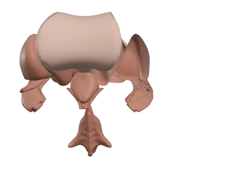
The brain ventricles are four connected cavities deep inside the brain. They are filled with cerebrospinal fluid (CSF), a clear liquid that cushions the brain and helps keep its chemical environment stable. Because the ventricles sit in the middle of the brain, learning them is also the easiest way to learn the deep structures packed around them: the thalamus, the basal nuclei and the hypothalamus.
Where it is
The ventricular system runs from the cerebral hemispheres down to the brainstem:
- The two lateral ventricles curve through the hemispheres, one on each side.
- Each lateral ventricle opens through a small interventricular foramen into the third ventricle, a narrow slit in the midline between the two halves of the thalamus.
- A thin channel through the midbrain, the cerebral aqueduct, leads down into the fourth ventricle, which lies between the brainstem in front and the cerebellum behind.
- From the fourth ventricle, CSF escapes through small openings into the space around the brain and spinal cord, and also continues into the narrow central canal of the spinal cord.
The ventricles are lined by a thin layer of cells called the ependyma.
Parts you can explore in the app
In the app you can open the ventricles on their own, with the structures around them, and study them part by part:
- Lateral ventricle (left) and Lateral ventricle (right): the largest ventricles, C-shaped cavities inside each cerebral hemisphere.
- Third ventricle: the thin midline cavity between the left and right thalamus.
- Fourth ventricle: the tent-shaped cavity between the brainstem and the cerebellum.
- Choroid plexus (left) and Choroid plexus (right): tufts of tiny blood vessels covered by ependymal cells that produce CSF. Choroid plexus is also found in the third and fourth ventricles.
- Corpus callosum: the broad band of white matter that links the two hemispheres and forms the roof of the lateral ventricles.
- Septum pellucidum: a thin membrane that hangs from the corpus callosum and separates the front parts of the two lateral ventricles.
- Fornix (left) and Fornix (right): arching bundles of nerve fibers that carry signals from the hippocampus toward the hypothalamus, involved in memory.
- Caudate nucleus (left) and Caudate nucleus (right): C-shaped nuclei whose head bulges into the wall of each lateral ventricle. They are part of the basal nuclei, which help regulate movement.
- Putamen (left) and Putamen (right): nuclei lying further out, which work with the caudate nucleus in controlling movement.
- Globus pallidus (left) and Globus pallidus (right): nuclei just inside the putamen that send much of the output of the basal nuclei.
- Thalamus (left) and Thalamus (right): egg-shaped relay stations that pass sensory and motor information to the cerebral cortex. They form the side walls of the third ventricle.
- Hypothalamus: the region below the thalamus that controls body temperature, hunger, thirst and the pituitary gland.
- Pineal gland: a small gland at the back of the third ventricle that releases melatonin.
Cross sections (sagittal, coronal and axial) are especially useful here, because the ventricles and these nuclei are deep and only show their shapes once the brain is cut.
What it does
The choroid plexus filters blood plasma and produces CSF. The fluid flows from the lateral ventricles to the third ventricle, through the cerebral aqueduct to the fourth ventricle, and then out into the subarachnoid space around the brain and spinal cord. It is finally absorbed through the arachnoid granulations into the large venous sinuses inside the dura mater, and so returns to the blood.
CSF does several things. It lets the brain float, which reduces its effective weight and protects it from knocks. It carries away waste products, and it keeps the fluid around neurons chemically stable. The cells of the choroid plexus also act as a barrier between the blood and the CSF.
The structures around the ventricles each have their own role:
- The basal nuclei (caudate nucleus, putamen and globus pallidus) help start wanted movements and suppress unwanted ones.
- The thalamus sorts and relays almost all sensory input on its way to the cortex.
- The hypothalamus keeps the internal environment stable and links the nervous system to the endocrine glands through the pituitary.
- The fornix is part of the circuits for memory.
- The pineal gland helps regulate daily sleep-wake rhythms through melatonin.
How to study it
- Draw the flow of CSF as a chain. Lateral ventricles, interventricular foramina, third ventricle, cerebral aqueduct, fourth ventricle, subarachnoid space, arachnoid granulations, venous blood. Exams ask for this order constantly.
- Name the walls. The corpus callosum is the roof of the lateral ventricles, the septum pellucidum their shared inner wall, and the caudate nucleus part of their outer wall. The thalamus forms the side walls of the third ventricle and the hypothalamus its floor.
- Learn the two groupings. Caudate nucleus + putamen = striatum. Putamen + globus pallidus = lentiform nucleus.
- Use the coronal section. It is the classic exam view: corpus callosum on top, lateral ventricles below it, the septum pellucidum between them, and the caudate, putamen and globus pallidus to the sides.
- Remember the lining. Ependymal cells line the ventricles; in the choroid plexus they make CSF.
Frequently asked questions
How many ventricles does the brain have?
Four: the two lateral ventricles, the third ventricle and the fourth ventricle.
Where is cerebrospinal fluid produced?
Mainly by the choroid plexus, which is found in all four ventricles.
What connects the third and fourth ventricles?
The cerebral aqueduct, a narrow channel that runs through the midbrain.
Where does cerebrospinal fluid go?
It flows out of the fourth ventricle into the subarachnoid space around the brain and spinal cord, and is absorbed into venous blood through the arachnoid granulations.