Quick answer
- Latin name
- Pulmones
- System
- Respiratory system
- Location
- Chest cavity, on either side of the heart, resting on the diaphragm
- Function
- Exchange oxygen and carbon dioxide between the air and the blood
- Parts in the app
- 8
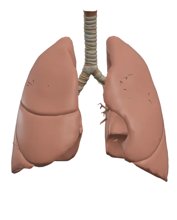
The lungs are two soft, spongy organs in the chest. Their job is simple to state: bring air and blood close enough together that oxygen can pass into the blood and carbon dioxide can pass out. To do this, the airway branches again and again, from the trachea down to millions of tiny air sacs.
Where it is
The lungs fill most of the chest cavity, one on each side of the heart. The ribs and the sternum protect them in front, and the spine behind.
- The apex of each lung rises just above the clavicle, into the root of the neck.
- The base is concave and rests on the dome of the diaphragm.
- On the inner surface, the hilum is where the main bronchus, the pulmonary artery, the pulmonary veins, nerves and lymphatic vessels enter or leave the lung.
Each lung sits in its own sealed pleural cavity. The visceral pleura covers the lung, the parietal pleura lines the chest wall, and a thin film of fluid between them lets the lung glide as you breathe.
The left lung is smaller than the right because the heart bulges into its side, leaving a hollow called the cardiac notch.
Parts you can explore in the app
In the app you can open the lungs on their own and study them part by part:
- Right upper lobe: the top lobe of the right lung, separated from the middle lobe by the horizontal fissure.
- Right middle lobe: a small wedge-shaped lobe found only on the right side.
- Right lower lobe: the large lower lobe, separated from the others by the oblique fissure.
- Left upper lobe: the top lobe of the left lung, including the lingula, a small tongue-shaped part below the cardiac notch.
- Left lower lobe: the lower lobe of the left lung, below the oblique fissure.
- Trachea: the windpipe, held open by C-shaped rings of cartilage, running from the larynx down into the chest.
- Left bronchial tree: the branching airways of the left lung, from the left main bronchus down to the smallest bronchioles.
- Right bronchial tree: the branching airways of the right lung, starting with a right main bronchus that is wider, shorter and more vertical than the left.
Cross sections (sagittal, coronal and axial) let you see the fissures between the lobes and follow the airways as they branch inside the lungs.
What it does
The airways fall into two zones.
The conducting zone moves air in and out: trachea, main bronchi, lobar bronchi, segmental bronchi, then smaller and smaller bronchioles down to the terminal bronchioles. Along the way the air is warmed, moistened and cleaned. Mucus traps dust and microbes, and tiny hairs called cilia sweep it up toward the throat.
The respiratory zone is where gas exchange happens: respiratory bronchioles, alveolar ducts and the alveoli, tiny air sacs wrapped in capillaries. Oxygen and carbon dioxide cross the very thin wall between air and blood by simple diffusion, each moving from where it is more concentrated to where it is less. Some cells in the alveolar wall make surfactant, a fluid that reduces surface tension and stops the alveoli from collapsing.
Breathing works by changing the volume of the chest. To breathe in, the diaphragm contracts and flattens and the external intercostal muscles lift the ribs. The chest gets bigger, the pressure inside the lungs drops below the pressure outside, and air flows in. Quiet breathing out is mostly passive: the muscles relax and the elastic lungs recoil. During heavy breathing, accessory muscles such as the sternocleidomastoid help lift the chest further.
The lungs receive the whole output of the right ventricle through the pulmonary arteries, which bring oxygen-poor blood for gas exchange. The lung tissue itself is fed by separate small bronchial arteries.
How to study it
- "Right has three, left has two." Three lobes and two fissures on the right; two lobes and one fissure on the left.
- Left lung landmarks. Cardiac notch and lingula. If you see them, you are looking at the left lung.
- The right bronchus is the "easy" path. Wider, shorter and more vertical, which is why inhaled objects more often end up in the right lung.
- Recite the branching order. Trachea, main bronchi, lobar bronchi, segmental bronchi, bronchioles, terminal bronchioles, respiratory bronchioles, alveolar ducts, alveoli.
- Connect pressure and volume. Bigger chest, lower pressure, air rushes in (Boyle's law). Many physiology questions come back to this idea.
- Use the axial section to see both lungs, the heart between them and the cardiac notch in one slice.
Frequently asked questions
How many lobes do the lungs have?
Five in total: three in the right lung (upper, middle and lower) and two in the left lung (upper and lower).
Why is the left lung smaller than the right?
The heart sits slightly to the left and takes up space, so the left lung has a cardiac notch and only two lobes.
Where does gas exchange happen?
In the alveoli, where oxygen moves from the air into the blood and carbon dioxide moves from the blood into the air.
Why do inhaled objects often go into the right lung?
The right main bronchus is wider, shorter and more vertical than the left, so it is the more direct path down from the trachea.