
Muscle · Upper limb
Extensor digitorum: origin, insertion, action and innervation
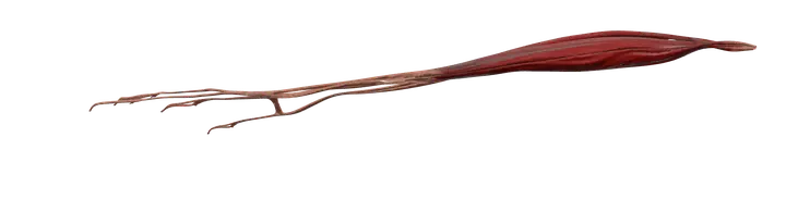
The extensor digitorum is a forearm muscle from the lateral epicondyle to the four fingers. It extends the fingers (posterior interosseous nerve).
Quick answer
- Latin name
- Musculus extensor digitorum
- Origin
- Lateral epicondyle of the humerus (common extensor origin)
- Insertion
- Extensor expansions of the index, middle, ring and little fingers, reaching the bases of the middle and distal phalanges
- Action
- Extends the four fingers, mainly at the knuckles (metacarpophalangeal joints); helps extend the wrist
- Innervation
- Posterior interosseous nerve, a branch of the radial nerve (C7–C8)
- Blood supply
- Posterior interosseous artery
See it on the 3D body
The extensor digitorum lit on the 3D forearm and hand, as in the forearm and hand quiz. Turn the body, peel the layers, tap any structure for its name.
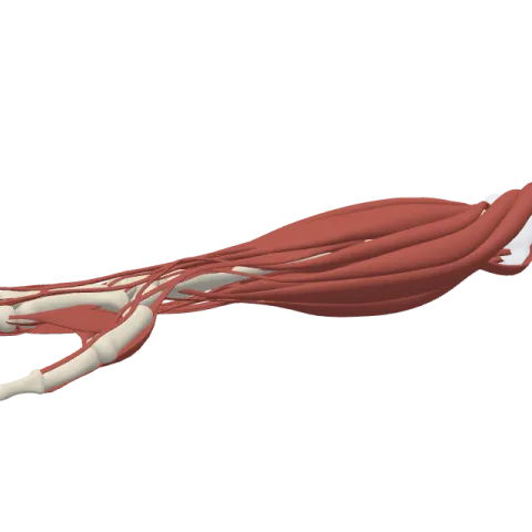
The extensor digitorum is the main muscle on the back of the forearm that straightens the index, middle, ring and little fingers. It runs from the lateral epicondyle of the humerus to the backs of the four fingers and is supplied by the posterior interosseous branch of the radial nerve.
Where it is
The extensor digitorum lies in the superficial layer of the posterior compartment of the forearm, the extensor side. It occupies the middle of the back of the forearm, between the extensor carpi radialis brevis on the thumb side and the extensor digiti minimi and extensor carpi ulnaris on the little-finger side. Its name is simple: extensor (straightens) digitorum (of the fingers). Older books call it the extensor digitorum communis.
Halfway down the forearm, the muscle divides into four tendons. They pass under the extensor retinaculum, a band of tough tissue across the back of the wrist, inside a shared sheath, together with the tendon of the extensor indicis. On the back of the hand they fan out, one to each finger. You can see them stand out under the skin when you spread your fingers and lift them.
Near the knuckles, the tendons are linked by small oblique bands called juncturae tendinum. These bands are why it is hard to lift the ring finger on its own while the other fingers stay curled: its tendon is tied to its neighbors.
Origin and insertion
- Origin: the lateral epicondyle of the humerus, through the common extensor tendon it shares with the extensor carpi radialis brevis, the extensor digiti minimi and the extensor carpi ulnaris.
- Insertion: each tendon spreads over the back of a finger into a triangular sheet called the extensor expansion (or dorsal hood).
- Central slip: the middle part of the expansion attaches to the base of the middle phalanx.
- Lateral bands: the two side parts rejoin over the finger and attach to the base of the distal phalanx.
The small muscles of the hand (lumbricals and interossei) also join the extensor expansion. See the bones of the hand for the phalanges and metacarpals.
What it does
- Extends the fingers at the knuckles: its strongest effect is at the metacarpophalangeal joints, lifting the four fingers back into line with the hand or beyond.
- Extends the finger joints: through the extensor expansion, it also straightens the two joints inside each finger (the proximal and distal interphalangeal joints). The lumbricals and interossei are essential for this part, especially when the knuckles are already straight.
- Helps extend the wrist: because its tendons cross the back of the wrist, it assists the wrist extensors (the two extensor carpi radialis muscles and the extensor carpi ulnaris).
You use it each time you open your hand to release an object, type, play the piano or flick something away. Two other muscles add independent movement: the extensor indicis for the index finger and the extensor digiti minimi for the little finger. That is why you can point with the index finger while the others stay bent.
Innervation and blood supply
The extensor digitorum is supplied by the posterior interosseous nerve (roots C7 and C8), the deep branch of the radial nerve. The radial nerve splits near the elbow; its deep branch winds around the radius through the supinator muscle and then supplies most of the muscles on the back of the forearm.
Its blood comes mainly from the posterior interosseous artery, which runs with the nerve.
How to remember it
- Lateral epicondyle = extensors, medial epicondyle = flexors. Think "Lateral Extensors" and "Medial Flexors".
- Radial nerve = all the extensors: in the arm and the forearm, every extensor muscle is supplied by the radial nerve or its branches.
- One muscle, four tendons: digitorum means "of the fingers", plural. The thumb has its own extensors.
- Central slip to the middle, lateral bands to the end: the expansion reaches both the middle and the distal phalanx.
The muscles of the forearm page shows the layers of the posterior compartment. The brachioradialis, also supplied by the radial nerve, lies on the thumb side of the same compartment.
Frequently asked questions
What is the action of the extensor digitorum?
It extends the index, middle, ring and little fingers, mainly at the knuckles, and helps extend the wrist.
Where does the extensor digitorum originate?
From the lateral epicondyle of the humerus, through the common extensor tendon.
Which nerve supplies the extensor digitorum?
The posterior interosseous nerve, the deep branch of the radial nerve, from roots C7 and C8.
Does the extensor digitorum extend the thumb?
No. The thumb has its own extensors, the extensor pollicis longus and brevis. The extensor digitorum only reaches the four fingers.
Why can't I lift my ring finger on its own?
Because its extensor tendon is linked to the tendons of the middle and little fingers by the juncturae tendinum, and it has no separate extensor muscle of its own.
