
Muscle · Upper limb
Abductor pollicis longus: origin, insertion, action and innervation
The abductor pollicis longus runs from the radius and ulna to the first metacarpal. It abducts and extends the thumb. Nerve: the posterior interosseous nerve.
Quick answer
- Latin name
- Musculus abductor pollicis longus
- Origin
- Posterior surfaces of the ulna and the radius, below the supinator, and the interosseous membrane between them
- Insertion
- Radial side of the base of the first metacarpal, often with a slip to the trapezium
- Action
- Abducts and extends the thumb at the carpometacarpal joint; helps abduct (radially deviate) the wrist
- Innervation
- Posterior interosseous nerve, from the deep branch of the radial nerve (C7-C8)
- Blood supply
- Posterior interosseous artery
See it on the 3D body
The abductor pollicis longus lit on the 3D forearm and hand, as in the forearm and hand quiz. Turn the body, peel the layers, tap any structure for its name.
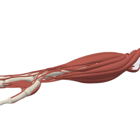
The abductor pollicis longus is a deep muscle on the back of the forearm whose tendon reaches the base of the thumb. It swings the thumb out to the side and back at the carpometacarpal joint, and it is supplied by the posterior interosseous nerve.
Where it is
The abductor pollicis longus (APL) belongs to the deep layer of the posterior compartment of the forearm, the extensor side. Four deep muscles in this layer serve the thumb and the index finger. From the thumb side to the little-finger side they are the APL, the extensor pollicis brevis, the extensor pollicis longus and the extensor indicis. The APL starts highest, just below the supinator.
In the lower third of the forearm, the APL and the extensor pollicis brevis come out from under the superficial extensors, between the extensor carpi radialis brevis and the extensor digitorum. Together they cross over the tendons of the two radial wrist extensors and slant toward the thumb. Anatomists call them "outcropping" muscles for this reason, like a layer of rock that surfaces in a hillside.
At the wrist, the two tendons share the first extensor compartment, a tunnel under the extensor retinaculum over the outer side of the radius. Below that, they form the front border of the anatomical snuffbox, the small hollow you see at the base of the thumb when you stretch it back.
Origin and insertion
- Origin: the back of the ulna and the radius, below the supinator, and the interosseous membrane, the sheet of fibrous tissue that joins the two bones.
- Insertion: the radial side of the base of the first metacarpal. A slip often reaches the trapezium, and the tendon is frequently split into two or more parts.
Note the level: the APL stops at the metacarpal, the long bone in the palm. Its short partner, the abductor pollicis brevis, goes one bone further, to the proximal phalanx. The metacarpals page shows the first metacarpal.
What it does
- Abduction of the thumb: at the carpometacarpal joint, it carries the first metacarpal away from the index finger, mostly to the side.
- Extension of the thumb: at the same joint, it pulls the thumb backward, working with the two thumb extensors.
- Help at the wrist: because its tendon crosses the thumb side of the wrist, it assists radial deviation, tilting the hand toward the thumb.
- Steadying the base of the thumb: it holds the first metacarpal in place while the short thumb muscles in the palm act on the rest of the thumb.
You use it when you open your hand wide to reach around something large, or when you stick the thumb out to hitch a ride.
Innervation and blood supply
Its nerve is the posterior interosseous nerve, carrying fibers from C7 and C8. That is the name the deep branch of the radial nerve takes once it has passed through the supinator into the back of the forearm.
Its blood comes from the posterior interosseous artery, which runs with the nerve between the superficial and deep layers.
De Quervain tenosynovitis is an irritation of the shared sheath of the APL and extensor pollicis brevis tendons in the first extensor compartment.
How to remember it
- APL and EPB travel as a pair: same layer, same compartment at the wrist, same border of the snuffbox.
- The long one stops short: the abductor pollicis longus ends on the first metacarpal, while the abductor pollicis brevis goes one bone further, to the proximal phalanx.
- One deep nerve for the deep layer: the four deep thumb and index muscles of the back of the forearm, the APL among them, all get the posterior interosseous nerve.
- The outcrop: picture two tendons surfacing between the wrist extensors and running diagonally to the thumb.
The muscles of the forearm page shows the superficial and deep extensors side by side. Then try to pick out the APL in the forearm and hand quiz.
Frequently asked questions
What is the action of the abductor pollicis longus?
It abducts and extends the thumb at the carpometacarpal joint and helps tilt the wrist toward the thumb.
Where does the abductor pollicis longus insert?
On the radial side of the base of the first metacarpal, often with a slip to the trapezium.
Which nerve supplies the abductor pollicis longus?
The posterior interosseous nerve (C7 and C8), which is the continuation of the deep branch of the radial nerve.
Which tendons form the anatomical snuffbox?
Two tendons run side by side along its front (palmar) edge: the APL and the extensor pollicis brevis. A single tendon, the extensor pollicis longus, bounds it at the back.
