Quick answer
- Latin name
- Cor
- System
- Cardiovascular system
- Location
- Middle of the chest, between the lungs and behind the sternum
- Function
- Pumps blood through the lungs and around the rest of the body
- Parts in the app
- 7
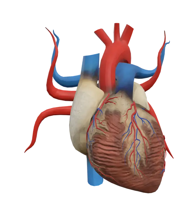
The heart is a hollow, muscular organ, roughly the size of a closed fist, that pumps blood without a break for an entire lifetime. It is really two pumps side by side: the right side sends blood to the lungs to pick up oxygen, and the left side sends that oxygen-rich blood to every other tissue of the body.
Where it is
The heart sits in the middle of the chest, in a space called the mediastinum, between the two lungs. It lies behind the sternum and the costal cartilages and rests on the diaphragm. The rib cage shields it, with large chest muscles such as the pectoralis major lying in front.
The heart is tilted. Its broad base, where the great vessels attach, faces up, back and to the right. Its pointed apex aims down, forward and to the left. This is why most of the heart lies a little to the left of the midline, even though it is a central organ.
A double-layered sac, the pericardium, wraps the heart. A thin film of fluid between its layers lets the heart beat with very little friction.
Parts you can explore in the app
In the app you can open the heart on its own and study it part by part:
- Heart: the whole organ, with its four chambers (two atria above, two ventricles below) and its thick muscular wall, the myocardium.
- Left atrium: receives oxygen-rich blood from the lungs through the pulmonary veins and passes it down to the left ventricle.
- Right atrium: receives oxygen-poor blood from the body through the superior and inferior venae cavae, and from the heart wall through the coronary sinus.
- Mitral valve: the valve between the left atrium and the left ventricle. It has two cusps, which is why it is also called the bicuspid valve.
- Tricuspid valve: the valve between the right atrium and the right ventricle, with three cusps.
- Coronary arteries: the vessels that feed the heart muscle itself. They arise from the base of the aorta, just above the aortic valve.
- Cardiac veins: the veins that drain the heart wall. Most of them empty into the coronary sinus, which opens into the right atrium.
Cross sections (sagittal, coronal and axial) let you look inside and compare the thin walls of the atria with the much thicker wall of the left ventricle.
What it does
Blood follows a single route through the heart, and the valves keep it moving forward:
- Oxygen-poor blood from the body enters the right atrium.
- It flows through the tricuspid valve into the right ventricle.
- The right ventricle pumps it through the pulmonary valve into the pulmonary trunk and on to the lungs, where it gives up carbon dioxide and takes up oxygen.
- Oxygen-rich blood returns through the pulmonary veins to the left atrium.
- It flows through the mitral valve into the left ventricle.
- The left ventricle pumps it through the aortic valve into the aorta and out to the whole body.
The tricuspid and mitral valves are the two atrioventricular valves. Thin cords called chordae tendineae tie their cusps to papillary muscles inside the ventricles. When the ventricles contract, these cords stop the cusps from flipping back into the atria. The closing of the atrioventricular valves, then of the pulmonary and aortic valves, makes the familiar "lub-dub" of the heartbeat.
The left ventricle has the thickest wall because it pushes blood through the long systemic circuit, while the right ventricle only has to reach the nearby lungs. Each beat starts in the sinoatrial node, a small cluster of specialized cells in the wall of the right atrium, so the heart sets its own rhythm. Nerves and hormones then speed it up or slow it down.
Like any muscle, the heart needs its own blood supply. The coronary arteries fill mostly while the heart muscle relaxes between beats, and the cardiac veins carry the used blood back to the right atrium.
How to study it
- Follow one drop of blood. Say the six steps above out loud until you can do them without notes. Many exam questions on the heart are really questions about this sequence.
- Name the valves with "LAB RAT". Left Atrioventricular = Bicuspid (mitral); Right Atrioventricular = Tricuspid.
- "Try before you buy." Blood meets the tricuspid valve before it meets the bicuspid (mitral) valve.
- Arteries leave, veins arrive. The pulmonary arteries carry oxygen-poor blood and the pulmonary veins carry oxygen-rich blood. Vessels are named by direction (away from or toward the heart), not by oxygen content. This is a favorite trick question.
- Learn the main coronary branches. The left coronary artery divides into the anterior interventricular artery (often called the left anterior descending) and the circumflex artery. The right coronary artery gives the marginal artery and, usually, the posterior interventricular artery. The great, middle and small cardiac veins drain into the coronary sinus.
- Use the cross sections. An axial slice shows both ventricles side by side and makes the difference in wall thickness obvious. A coronal slice shows each atrium sitting above its ventricle, with the valve between them.
Frequently asked questions
How many chambers does the heart have?
Four: the right and left atria, and the right and left ventricles. A muscular wall, the septum, separates the right side of the heart from the left.
What is the difference between the mitral and tricuspid valves?
Both are atrioventricular valves. The mitral valve sits on the left side and has two cusps; the tricuspid valve sits on the right side and has three.
Where do the coronary arteries come from?
The right and left coronary arteries branch from the aorta, just above the aortic valve. They are the first branches of the aorta.
Is the heart on the left side of the chest?
It sits near the middle of the chest, behind the sternum. Its apex points to the left, so more of the heart lies left of the midline than right of it.