Table · Trunk
Muscles of the back: superficial, intermediate and deep layers
The muscles of the back in three layers: trapezius, latissimus dorsi and rhomboids on the surface, the erector spinae and the deep muscles along the spine.
The muscles of the back lie in three layers. The superficial layer (trapezius, latissimus dorsi, levator scapulae and the two rhomboids) runs from the skull and the spine to the clavicle, the scapula or the humerus and moves the shoulder and the arm. The intermediate layer is two thin muscles on the ribs, the serratus posterior superior and inferior. The deep layer runs along the spine itself: the splenius muscles, the erector spinae (iliocostalis, longissimus, spinalis) and, under it, the short transversospinal muscles. The deep layer holds the spine upright, extends it, bends it to the side and turns it.
At a glance
| Muscle | Origin | Insertion | Action |
|---|---|---|---|
| Trapezius | Occipital bone, Nuchal ligament, Vertebrae C7–T12 | Clavicle, Scapula | Elevates, retracts and depresses the scapula; rotates it upward |
| Latissimus dorsi | Vertebrae T7–L5, Sacrum, Hip bone, Ribs 9–12 | Humerus | Extends, adducts and medially rotates the arm |
| Levator scapulae | Atlas (C1), Axis (C2), Vertebra C3, Vertebra C4 | Scapula | Elevates the scapula |
| Rhomboid major | Vertebrae T2–T5 | Scapula | Retracts and elevates the scapula |
| Rhomboid minor | Vertebra C7, Vertebra T1 | Scapula | Retracts and elevates the scapula |
| Iliocostalis | Sacrum, Hip bone, Ribs | Ribs, Cervical vertebrae | Extends and laterally flexes the vertebral column |
| Longissimus | Sacrum, Lumbar and thoracic vertebrae | Ribs, Thoracic and cervical vertebrae, Temporal bone | Extends and laterally flexes the vertebral column and head |
| Spinalis | Spinous processes of upper lumbar and lower thoracic vertebrae | Spinous processes of upper thoracic vertebrae | Extends the vertebral column |
| Quadratus lumborum | Hip bone | Twelfth rib, Vertebrae L1–L4 | Laterally flexes the trunk; steadies the twelfth rib |
From the anatomical data behind Anatomy Atlas 3D (Z-Anatomy, CC BY-SA 4.0, reviewed). Names link to the detailed page when there is one.
How they are organized
All three layers start from the vertebral column, so the bones of the spine are the map to read them on. What separates the layers is where the muscles end and which nerves supply them. Nine of these muscles are in the app and in the table above, with their attachments. Each muscle named below is linked when it has a page of its own.
Superficial layer: from the spine to the shoulder
These five muscles sit on the back but work on the upper limb, which is why they are counted among the extrinsic back muscles.
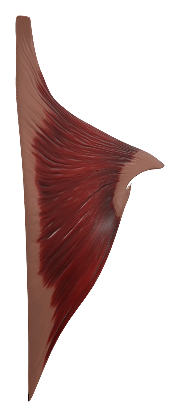
- The trapezius is the large triangular muscle of the upper back; the right and left ones together form a kite. It runs from the occipital bone and the spinous processes down to T12 out to the clavicle and the scapula.
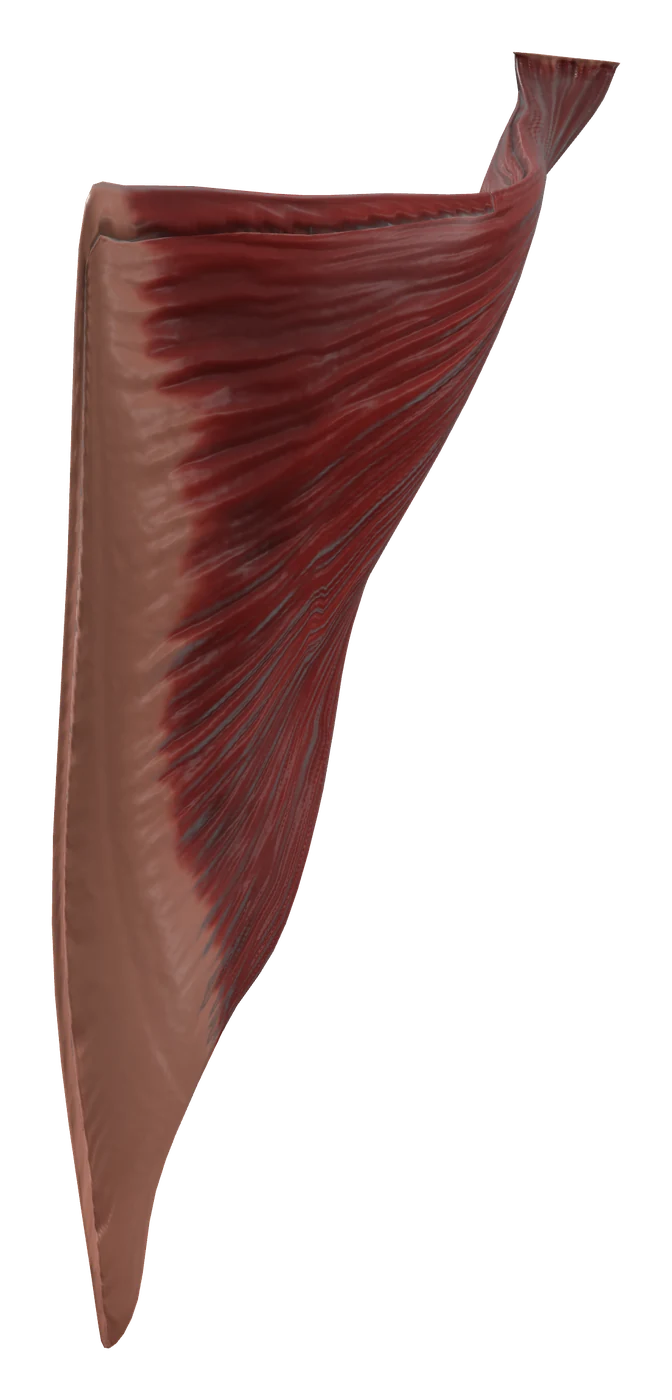
- The latissimus dorsi covers the lower back. It starts on the lower thoracic vertebrae, the thoracolumbar fascia, the iliac crest and the lower ribs, and ends on the front of the humerus.
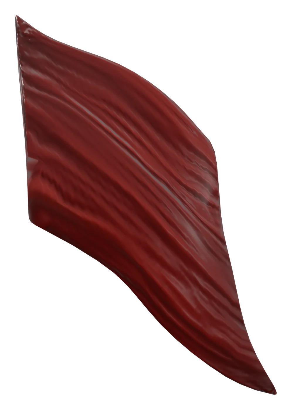
- Three muscles go to the medial border of the scapula, the rhomboids under the trapezius and the levator scapulae partly under it: the levator scapulae from the upper four cervical vertebrae, then the rhomboid minor from C7 and T1 and the rhomboid major from T2 to T5.
The muscles on the scapula itself (supraspinatus, infraspinatus, teres minor and teres major) are seen from the back too, but they move the shoulder joint and belong to the muscles of the shoulder.
Intermediate layer: two muscles on the ribs
The serratus posterior superior runs from C7 and the upper thoracic spinous processes down to ribs 2 to 5, and the serratus posterior inferior from the lower thoracic and upper lumbar spinous processes up to the last four ribs. Both are thin sheets, traditionally classed with the muscles of breathing because they end on the ribs. They are not among the nine muscles in the table.
Deep layer: the muscles of the spine
These are the intrinsic or "true" back muscles. They lie in the groove on each side of the spinous processes, under the thoracolumbar fascia.
- Splenius muscles, at the back of the neck: the splenius capitis and the splenius cervicis wrap over the deeper muscles of the neck. They are described with the muscles of the neck.
- Erector spinae, three columns that run from the sacrum and the iliac crest up to the ribs, the vertebrae and the skull. From the side toward the midline: the iliocostalis, the longissimus and the spinalis.
- Transversospinal muscles, under the erector spinae: bundles that run obliquely upward from the transverse processes toward the midline, long in the semispinalis, shorter in the multifidus, shortest in the rotatores. The group is the semispinalis (semispinalis capitis and semispinalis cervicis in the neck), the multifidus and the rotatores.
The quadratus lumborum lies in front of the erector spinae, between the twelfth rib and the iliac crest. Anatomists class it with the posterior abdominal wall, so it also appears in the table of the abdominal muscles, but it is listed here because it fills the lower back between the last rib and the pelvis.
What they do together
- Moving the scapula. The upper trapezius and the levator scapulae lift it (a shrug). The middle trapezius and the rhomboids pull it toward the spine. The lower trapezius pulls it down, and the upper and lower trapezius together turn the socket of the shoulder upward when the arm is raised.
- Moving the arm. The latissimus dorsi extends the arm, pulls it toward the body and rotates it medially. With the arms fixed overhead, the same muscle pulls the trunk up toward them, as in climbing or a pull-up.
- Holding the spine upright and extending it. When both sides contract, the erector spinae extends the vertebral column. It also works while you bend forward, lengthening slowly to control the movement, and then pulls the trunk back up.
- Bending to the side. The erector spinae and the quadratus lumborum of one side bend the trunk toward that side.
- Turning the trunk. The transversospinal muscles of one side turn the trunk toward the opposite side, working with the oblique abdominal muscles.
- Breathing. The quadratus lumborum steadies the twelfth rib so that the diaphragm has a fixed edge to pull on.
How to learn them
- Go layer by layer. Ask of each muscle where it goes. To the clavicle, the scapula or the humerus: superficial layer. A thin sheet from the spine to the ribs: intermediate. Up along the spine itself: deep layer. The quadratus lumborum is the exception, a muscle of the abdominal wall behind the others.
- "I Love Spine" for the erector spinae. From lateral to medial: Iliocostalis, Longissimus, Spinalis. The names say where each column goes: ilio-costalis from the ilium to the ribs, longissimus the longest, spinalis between spinous processes.
- Rhomboids by their vertebrae. Minor above (C7 and T1), major below (T2 to T5). Both end on the medial border of the scapula and both are supplied by the dorsal scapular nerve.
- Nerves by layer. The deep muscles are supplied by the posterior (dorsal) rami of the spinal nerves, segment by segment. The superficial muscles get nerves that come from the neck and the brachial plexus: the accessory nerve (cranial nerve XI) for the trapezius, the dorsal scapular nerve for the rhomboids and part of the levator scapulae, the thoracodorsal nerve for the latissimus dorsi. The two serratus posterior muscles get intercostal nerves, and the quadratus lumborum the anterior rami of T12 to L4. The spinal levels and the arteries are on the page of each muscle.
- One gap to know. The lower edge of the trapezius, the upper edge of the latissimus dorsi and the medial border of the scapula frame a small gap called the triangle of auscultation.
In the app, peel the back one layer at a time: with the trapezius and the latissimus dorsi hidden, the rhomboids and the three columns of the erector spinae appear, and each column can be followed from the sacrum up to the neck. Test yourself with the free back quiz and the trunk muscles quiz, and go on with the lumbar vertebrae and the thoracic vertebrae the deep muscles attach to.
Frequently asked questions
What are the main muscles of the back?
On the surface, the trapezius, the latissimus dorsi, the levator scapulae and the rhomboid major and minor. Deep along the spine, the erector spinae (iliocostalis, longissimus, spinalis), the splenius muscles and the transversospinal muscles (semispinalis, multifidus, rotatores).
What are the three erector spinae muscles?
The iliocostalis, the longissimus and the spinalis, in that order from the side toward the midline. Together they extend the spine, and those of one side bend it to that side.
What is the largest muscle of the back?
The latissimus dorsi is the widest: its name means "widest of the back". It covers the lower half of the back and ends on the humerus, so its job is to move the arm.
Which nerves supply the back muscles?
The deep (intrinsic) back muscles are supplied by the posterior rami of the spinal nerves. The superficial muscles are supplied by other nerves because they move the upper limb: the accessory nerve for the trapezius, the dorsal scapular nerve for the rhomboids, the thoracodorsal nerve for the latissimus dorsi.
What is the difference between extrinsic and intrinsic back muscles?
Extrinsic muscles (the superficial and intermediate layers) lie on the back but move the shoulder, the arm or the ribs. Intrinsic muscles (the deep layer) run between the pelvis, the spine, the ribs and the skull, move the spine itself and are supplied by the posterior rami.
Detailed pages
 Trapezius
The diamond-shaped muscle that moves and steadies the shoulder blade.
 Latissimus dorsi
The broadest muscle of the back, from the spine and pelvis to the humerus.
Levator scapulae
The strap-like neck muscle that lifts the shoulder blade.
 Rhomboid major
The flat muscle that pulls the shoulder blade toward the spine.
Rhomboid minor
The narrow band above the rhomboid major that pulls the shoulder blade toward the spine.
Iliocostalis
The outer column of the erector spinae, climbing the angles of the ribs.
Longissimus
The middle and largest column of the erector spinae, reaching the skull.
Spinalis
The thin inner column of the erector spinae, from spine to spine.
Quadratus lumborum
The deep square muscle of the lower back, between the 12th rib and the pelvis.
Sources
- OpenStax, Anatomy and Physiology 2e, 11.3 Axial muscles of the head, neck and back
- OpenStax, Anatomy and Physiology 2e, 11.5 Muscles of the pectoral girdle and upper limbs
- OpenStax, Anatomy and Physiology 2e, 11.4 Axial muscles of the abdominal wall and thorax
- OpenStax, Anatomy and Physiology 2e, 13.4 The peripheral nervous system
- OpenStax, Anatomy and Physiology 2e, 7.3 The vertebral column
- Gray, Anatomy of the Human Body (1918), The muscles connecting the upper extremity to the vertebral column
- Gray, Anatomy of the Human Body (1918), The deep muscles of the back
- Gray, Anatomy of the Human Body (1918), The muscles of the thorax
- Z-Anatomy, the libre 3D atlas of anatomy
