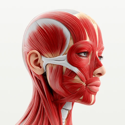
Table · Head and neck
Muscles of the neck: sternocleidomastoid, scalenes and hyoid muscles
The muscles of the neck: the sternocleidomastoid, the three scalenes, the muscles above and below the hyoid bone, and the back muscles that hold the head up.
The main muscles of the neck fall into four groups: the sternocleidomastoid on each side, the hyoid muscles at the front (above and below the hyoid bone), the three scalenes deep at the side, and the muscles of the back of the neck, such as the splenius, the levator scapulae and the upper trapezius. Together they turn and hold up the head, move the hyoid bone and larynx in swallowing, and lift the first two ribs in a deep breath. Two deep sets are not covered here: the prevertebral muscles on the front of the spine (longus colli and longus capitis) and the small suboccipital muscles under the skull.
At a glance
| Muscle | Origin | Insertion | Action |
|---|---|---|---|
| Sternocleidomastoid | Manubrium of sternum, Clavicle | Temporal bone | Turns the head to the opposite side; flexes the neck |
| Scalenus anterior | Vertebrae C3–C6 | First rib | Elevates the first rib; flexes the neck |
| Scalenus medius | Vertebrae C2–C7 | First rib | Elevates the first rib; bends the neck to the side |
| Scalenus posterior | Vertebrae C4–C6 | Second rib | Elevates the second rib; bends the neck to the side |
| Mylohyoid | Mandible | – | Raises the floor of the mouth and the hyoid; depresses the mandible |
| Thyrohyoid | Thyroid cartilage | Hyoid bone | Depresses the hyoid; raises the larynx |
| Splenius cervicis | – | Atlas (C1), Axis (C2) | Extends and rotates the neck to the same side |
| Levator scapulae | Atlas (C1), Axis (C2), Vertebra C3, Vertebra C4 | Scapula | Elevates the scapula |
| Trapezius | Occipital bone, Nuchal ligament, Vertebrae C7–T12 | Clavicle, Scapula | Elevates, retracts and depresses the scapula; rotates it upward |
From the anatomical data behind Anatomy Atlas 3D (Z-Anatomy, CC BY-SA 4.0, reviewed). Names link to the detailed page when there is one.
How they are organized
The neck is built around the seven cervical vertebrae. The airway and the esophagus run in front of them, and the muscles are arranged around both. The table above lists nine of them; the text below links every neck muscle that has its own page.
The sternocleidomastoid, the landmark
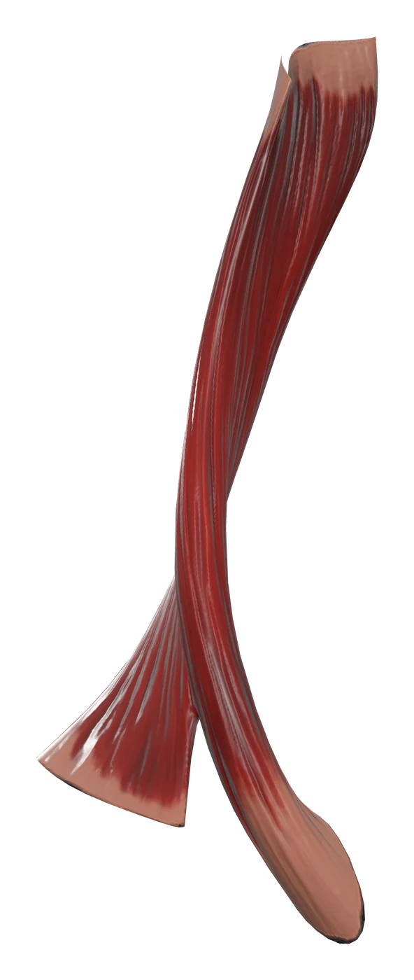
The sternocleidomastoid runs from the sternum and the clavicle up to the mastoid process behind the ear. It crosses the side of the neck diagonally and splits it into two triangles: the anterior triangle in front of it, which holds the hyoid muscles, and the posterior triangle behind it, whose floor is made by the splenius capitis, the levator scapulae and the middle and posterior scalenes. Over all of this, just under the skin, lies the thin platysma.
The hyoid muscles, at the front
The hyoid is a small U-shaped bone under the chin that touches no other bone. Two sets of muscles hold it.
- Suprahyoid muscles, between the hyoid and the skull or the mandible: the mylohyoid, which forms the floor of the mouth, the digastric, the stylohyoid and the geniohyoid.
- Infrahyoid muscles, the four "strap" muscles below the hyoid: the sternohyoid and the omohyoid on the surface, the sternothyroid and the thyrohyoid underneath, lying on the larynx and the thyroid gland.
The scalenes, deep at the side
The anterior, middle and posterior scalene run from the transverse processes of the cervical vertebrae down to the ribs: the anterior and middle to the first rib, the posterior to the second. The roots of the brachial plexus and the subclavian artery pass through the gap between the anterior and the middle scalene, on their way to the arm.
The back of the neck
From the surface inwards: the upper trapezius, then the levator scapulae and the two splenius muscles (splenius capitis to the skull, splenius cervicis to the upper cervical vertebrae), then the semispinalis capitis. The trapezius ends on the clavicle and the scapula and the levator scapulae on the scapula, so they also belong to the muscles of the shoulder.
What they do together
- Turning the head. One sternocleidomastoid turns the face to the opposite side. The splenius muscles turn it to their own side. To look left, the right sternocleidomastoid and the left splenius work together; about half of the movement happens at the atlanto-axial joint.
- Bending forward and holding the head up. Both sternocleidomastoids and the scalenes flex the neck. The splenius, the semispinalis capitis and the upper trapezius extend it, and they work all day because the center of gravity of the head lies in front of its joint with the spine.
- Tilting to the side. The scalenes, the sternocleidomastoid, the levator scapulae and the splenius of the same side bend the neck toward the shoulder.
- Swallowing and speaking. The suprahyoid muscles lift the hyoid and the floor of the mouth when you swallow, and the thyrohyoid pulls the larynx up toward the hyoid. Afterwards the sternohyoid, the omohyoid and the sternothyroid pull the hyoid and the larynx back down. With the hyoid held still from below, the digastric and the mylohyoid help open the mouth.
- Breathing in deeply. With the head and neck held still, the scalenes lift the first two ribs and the sternocleidomastoid lifts the sternum. They are called accessory muscles of inspiration because quiet breathing is done mostly by the diaphragm.
How to learn them
- Start from the sternocleidomastoid. Find it first, then place every other muscle in front of it (hyoid muscles), behind it (scalenes, levator scapulae, splenius) or under it.
- Read the names of the hyoid muscles. Most names give the two attachments, origin first: sterno-hyoid (sternum to hyoid), sterno-thyroid (sternum to thyroid cartilage), thyro-hyoid (thyroid cartilage to hyoid), omo-hyoid (omo means shoulder: scapula to hyoid), mylo-hyoid (the molar region of the mandible to hyoid), stylo-hyoid (styloid process to hyoid). The digastric is the exception: its name means "two bellies".
- Scalenes by their rib. Anterior and middle end on the first rib, posterior on the second. The plexus and the artery pass between the anterior and the middle.
- Nerves by group. The accessory nerve (cranial nerve XI) supplies the sternocleidomastoid and the trapezius. The ansa cervicalis (C1 to C3) supplies the sternohyoid, the omohyoid and the sternothyroid; the thyrohyoid gets C1 fibers that travel with the hypoglossal nerve. The scalenes get branches of the anterior rami of the cervical nerves, the splenius muscles branches of the posterior rami. The mylohyoid is supplied by the mandibular nerve (V3), the stylohyoid and the platysma by the facial nerve (VII), and the digastric by both, one belly each. Each muscle's page gives its nerve and blood supply in detail.
In the app, hide the platysma and the sternocleidomastoid to uncover the strap muscles and the scalenes, isolate one muscle and turn the model to follow it from one attachment to the other. Then test yourself with the free head and neck muscles quiz, and go on with the muscles of the face, which share the same region in most courses.
Frequently asked questions
What are the main muscles of the neck?
The sternocleidomastoid, the three scalenes, the suprahyoid and infrahyoid muscles at the front, and at the back the splenius muscles, the levator scapulae, the semispinalis capitis and the upper trapezius. The platysma covers the front and sides under the skin.
Which muscle turns the head?
The sternocleidomastoid turns the face to the opposite side: the right one turns the head to the left. The splenius muscles of the side you turn toward help it.
What are the infrahyoid (strap) muscles?
Four flat muscles below the hyoid bone: sternohyoid, omohyoid, sternothyroid and thyrohyoid. They pull the hyoid and the larynx down after swallowing. The thyrohyoid is the odd one: it lowers the hyoid but raises the larynx when the hyoid is held still.
What passes between the scalene muscles?
The roots of the brachial plexus and the subclavian artery pass between the anterior and the middle scalene. The subclavian vein passes in front of the anterior scalene.
Is the trapezius a neck muscle or a back muscle?
Both. Its upper fibers start on the occipital bone and the nuchal ligament and form the outline of the back of the neck, while the rest covers the upper back. It moves the scapula, so it is usually listed with the shoulder muscles as well.
Detailed pages
 Sternocleidomastoid
The strap of the neck that turns the head to the other side.
Anterior scalene
The deep neck muscle that lifts the first rib and guides the phrenic nerve.
Middle scalene
The largest scalene, behind the brachial plexus, lifting the first rib.
Posterior scalene
The smallest scalene, the only one that reaches the second rib.
Mylohyoid
The muscular sheet that forms the floor of the mouth.
Thyrohyoid
The short strap muscle between the larynx and the hyoid.
Splenius cervicis
The lower splenius muscle, pulling on the atlas and the axis.
Levator scapulae
The strap-like neck muscle that lifts the shoulder blade.
 Trapezius
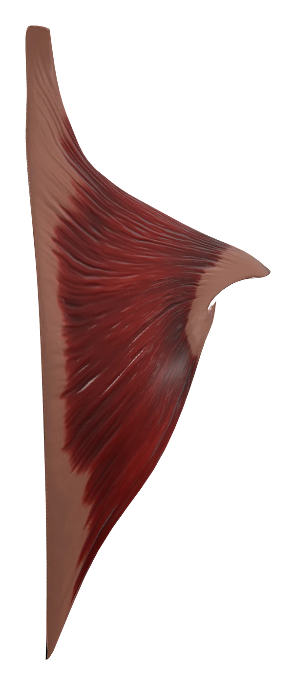
The diamond-shaped muscle that moves and steadies the shoulder blade.
Sources
- OpenStax, Anatomy and Physiology 2e, 11.3 Axial muscles of the head, neck and back
- OpenStax, Anatomy and Physiology 2e, 11.5 Muscles of the pectoral girdle and upper limbs
- OpenStax, Anatomy and Physiology 2e, 13.4 The peripheral nervous system
- OpenStax, Anatomy and Physiology 2e, 7.3 The vertebral column
- Z-Anatomy, the libre 3D atlas of anatomy
