
Muscle · Head and neck
Masseter muscle: origin, insertion, action and innervation
The masseter runs from the zygomatic arch to the angle and ramus of the mandible. It closes the jaw and is supplied by the masseteric nerve from V3.
Quick answer
- Latin name
- Musculus masseter
- Origin
- Lower border and deep surface of the zygomatic arch of the skull, with the zygomatic process of the maxilla
- Insertion
- Lateral surface of the ramus and angle of the mandible, up to the coronoid process
- Action
- Elevates the mandible (closes the jaw); superficial fibers help protrude it, deep fibers help retract it
- Innervation
- Masseteric nerve, from the mandibular division of the trigeminal nerve (V3)
- Blood supply
- Masseteric artery (from the maxillary artery), with branches of the facial and transverse facial arteries
See it on the 3D body
The masseter lit on the 3D head, as in the head quiz. Turn the body, peel the layers, tap any structure for its name.
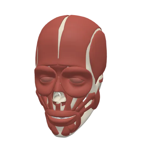
The masseter is the thick, four-sided chewing muscle on the side of the face, running from the cheekbone down to the angle of the lower jaw. It closes the mouth and presses the teeth together, and it is supplied by the masseteric nerve, a branch of the mandibular division of the trigeminal nerve (V3).
Where it is
The masseter covers the outer surface of the ramus of the mandible, the vertical part of the lower jaw between the ear and the cheek. Its upper edge hangs from the zygomatic arch, the bar of bone you can feel running from the cheek back toward the ear. Its lower edge reaches the angle of the mandible, the corner of the jaw below the earlobe.
To find it, put your fingers on your cheek in front of the earlobe and clench your teeth: the muscle bulges and turns hard.
A few structures lie on or around it:
- The back part of the muscle is covered by the parotid gland, the largest salivary gland.
- The parotid duct crosses its outer surface, then turns inward at its front edge to open into the mouth.
The masseter is one of the four muscles of mastication, with the temporalis and the medial and lateral pterygoids.
Origin and insertion
The masseter has two layers, and the app shows them as two separate structures, the superficial part and the deep part.
- Superficial part: arises by a strong tendon from the front two thirds of the lower border of the zygomatic arch, including the zygomatic process of the maxilla. Its fibers run downward and backward to the angle and the lower half of the outer surface of the ramus.
- Deep part: smaller, it arises from the back third and the inner surface of the zygomatic arch. Its fibers run almost straight down to the upper half of the ramus and the outer side of the coronoid process.
The zygomatic arch is formed by the zygomatic and temporal bones of the skull; both layers end on the mandible.
What it does
- Closing the jaw: the masseter lifts the mandible at the temporomandibular joint and brings the lower teeth against the upper teeth. It does most of the work when you bite hard.
- Small forward and backward movements: the slanted superficial fibers help push the jaw forward (protrusion), and the more vertical deep fibers help pull it back (retraction).
- Grinding: working with the pterygoid muscles, it helps move the jaw during the side-to-side grinding of chewing.
Opening the mouth is done by other muscles, mainly the lateral pterygoid and the muscles under the chin.
Innervation and blood supply
The masseter is supplied by the masseteric nerve, a branch of the mandibular division of the trigeminal nerve (V3), cranial nerve V. The nerve reaches the deep surface of the muscle through the mandibular notch, the dip between the coronoid process and the condyle. The expression muscles of the face, by contrast, are supplied by the facial nerve.
Its main blood supply is the masseteric artery, a branch of the maxillary artery that travels with the nerve through the mandibular notch. Branches of the facial artery and the transverse facial artery add to it.
How to remember it
- Masseter sounds like "mastication": both come from Greek words for chewing.
- Feel it, then name it: the muscle that bulges in front of the earlobe when you clench is the masseter. The one at the temple is the temporalis.
- Two layers, two directions: superficial fibers slant back and help push the jaw forward, deep fibers drop straight down and help pull it back.
- Arch to angle: zygomatic arch above, angle of the mandible below.
The muscles of the face page places the masseter among the other face muscles. Test yourself in the head quiz.
Frequently asked questions
What is the main action of the masseter?
It elevates the mandible, which closes the jaw and clenches the teeth together.
Where does the masseter originate and insert?
It starts on the zygomatic arch and inserts on the outer surface of the ramus and angle of the mandible.
Which nerve supplies the masseter?
The masseteric nerve, a branch of the mandibular division of the trigeminal nerve (V3).
Is the masseter a muscle of facial expression?
No. It is a muscle of mastication, supplied by the trigeminal nerve. The muscles of facial expression are supplied by the facial nerve.
What is the difference between the masseter and the temporalis?
Both close the jaw. The masseter lies on the side of the mandible below the zygomatic arch, while the temporalis covers the temple above the arch and ends on the coronoid process.
