
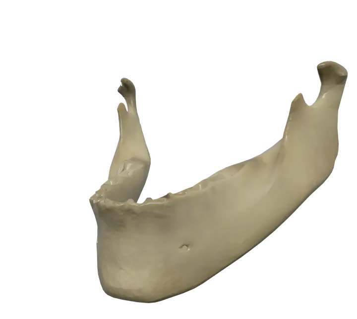
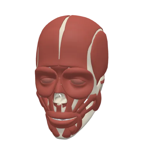
See it on the 3D body
The mandible lit on the 3D head, as in the head quiz. Turn the body, peel the layers, tap any structure for its name.
Quick answer
- Latin name
- Mandibula
- Type
- Irregular bone
- Location
- Lower face, forming the lower jaw and the chin
- Joints
- Temporomandibular joint with the temporal bone on each side; the lower teeth sit in its sockets (gomphoses)
The mandible is the bone of the lower jaw: the largest and strongest bone of the face, and the only bone of the skull that moves freely, at the temporomandibular joints. It holds the lower teeth, forms the chin and gives attachment to the chewing muscles.
Overall shape
The mandible is shaped like a horseshoe with two upright arms. It has three main parts:
- The body: the curved, horizontal part that holds the lower teeth and forms the chin.
- Two rami (singular: ramus): the flat, vertical plates that rise from the back of the body on each side, in front of the ears.
- The angle: the corner where the lower border of the body meets the back of the ramus, on each side. You can feel it below and in front of the earlobe.
At birth the mandible is made of two halves, joined in the midline at the mandibular symphysis. They fuse into a single bone during the first year of life.
Body
- The alveolar process is the upper edge of the body, with sockets for the 16 lower teeth in adults. Each tooth is held in its socket by a fibrous joint called a gomphosis.
- The mental protuberance is the triangular bump at the front that forms the chin.
- The mental foramen is a small opening on the outer surface, usually below the second premolar tooth. The mental nerve and vessels come out through it to supply the skin of the chin and lower lip.
- On the inner surface, small bumps in the midline called the mental spines (genial tubercles) anchor the genioglossus (a muscle of the tongue) and the geniohyoid.
- Just beside them, the digastric fossa receives the anterior belly of the digastric muscle.
- The mylohyoid line, an oblique ridge on the inner surface, anchors the mylohyoid muscle, which forms the floor of the mouth.
Ramus and processes
- The coronoid process is the thin, pointed process at the front of the top of the ramus. The temporalis inserts on it.
- The condylar process is at the back. Its rounded head (condyle) fits into the mandibular fossa of the temporal bone to form the jaw joint. Just below the head, the narrower neck has a small pit in front, the pterygoid fovea, for the lateral pterygoid muscle.
- The mandibular notch is the U-shaped dip between the two processes.
- On the inner surface of the ramus, the mandibular foramen opens into a canal that runs through the body. The inferior alveolar nerve and vessels enter here to supply the lower teeth. A small tongue of bone, the lingula, partly covers the opening.
Joints
The mandible forms the temporomandibular joint (TMJ) on each side, with the mandibular fossa and articular tubercle of the temporal bone. The joint sits just in front of the external opening of the ear: put a finger there and open your mouth to feel the condyle move.
The TMJ is a synovial joint with an articular disc of fibrocartilage that divides it into an upper and a lower compartment. This allows two kinds of movement: a hinge movement in the lower compartment and a forward gliding movement in the upper compartment. When you open your mouth wide, the condyle both rotates and slides forward onto the articular tubercle. The two joints always work together, because the mandible is a single bone.
Muscles that attach to it
- Muscles of mastication: the temporalis on the coronoid process; the masseter on the outer surface of the ramus and angle; the medial pterygoid on the inner surface of the ramus and angle; the lateral pterygoid on the neck of the condyle and the joint disc.
- Muscles under the chin: the mylohyoid, the geniohyoid and the anterior belly of the digastric, which help open the mouth and support the floor of the mouth.
- Tongue: the genioglossus, from the mental spines.
- Muscles of facial expression: the buccinator, the depressor anguli oris, the depressor labii inferioris and the mentalis, plus the platysma along the lower border. The muscles of the face page describes them.
Chewing is the first step of digestion, described on the stomach and intestines page.
Frequently asked questions
Is the mandible the only movable bone of the skull?
Yes, apart from the tiny ossicles of the middle ear. It is the only skull bone that moves at a true joint with the rest of the skull, the temporomandibular joint.
What is the difference between the mandible and the maxilla?
The mandible is the lower jaw and moves. The maxilla is the upper jaw; it is fixed to the rest of the skull and holds the upper teeth.
What attaches to the coronoid process of the mandible?
The temporalis muscle, which closes the jaw.
What passes through the mental foramen?
The mental nerve and vessels, which supply the skin of the chin and the lower lip.
What joint does the mandible form?
The temporomandibular joint, on each side, with the temporal bone. It is a synovial joint with an articular disc.
Muscles that originate here
Muscles that insert here
- Deep part of masseter
- Superficial part of masseter
- Temporalis
From the anatomical data behind Anatomy Atlas 3D (Z-Anatomy, reviewed). In the app, tap the bone, then each muscle around it to open its card.
