
Muscle · Head and neck
Temporalis: origin, insertion, action and innervation
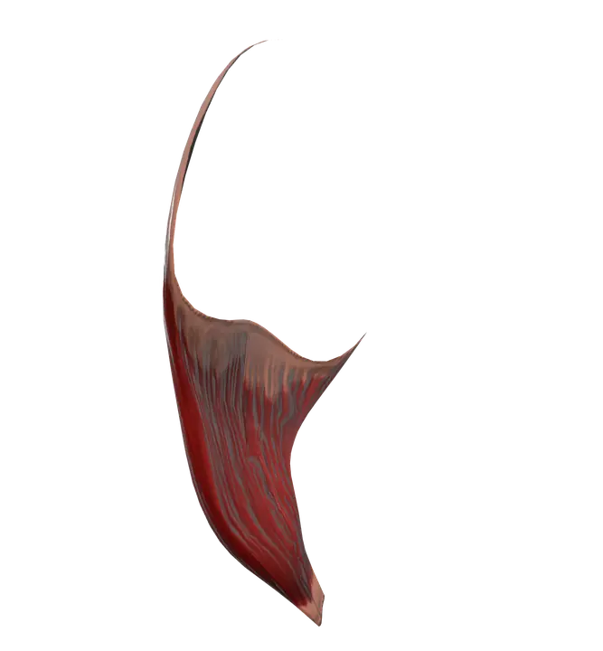
The temporalis is a fan-shaped chewing muscle from the temporal fossa to the coronoid process of the mandible. It closes the jaw (mandibular nerve, V3).
Quick answer
- Latin name
- Musculus temporalis
- Origin
- Floor of the temporal fossa (frontal, parietal, temporal and sphenoid bones) and the deep surface of the temporal fascia
- Insertion
- Coronoid process and front edge of the ramus of the mandible
- Action
- Elevates the mandible (closes the jaw); its posterior fibers retract it; helps side-to-side grinding
- Innervation
- Deep temporal nerves, from the mandibular division of the trigeminal nerve (V3)
- Blood supply
- Deep temporal arteries (from the maxillary artery) and the middle temporal artery (from the superficial temporal artery)
See it on the 3D body
The temporalis lit on the 3D head, as in the head quiz. Turn the body, peel the layers, tap any structure for its name.
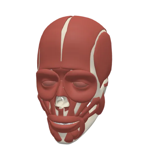
The temporalis is a broad, fan-shaped chewing muscle on the side of the head, at the temple, running from the temporal fossa of the skull to the coronoid process of the mandible. It closes the jaw and pulls it backward, and it is supplied by the mandibular branch of the trigeminal nerve.
Where it is
The temporalis covers the temporal fossa, the wide, shallow area on the side of the skull above the cheekbone. The fossa is bounded above by a curved ridge, the temporal line, and below by the zygomatic arch, the bony bridge you can feel between the cheek and the ear. The muscle is covered by a strong sheet of connective tissue, the temporal fascia, which stretches from the temporal line down to the zygomatic arch.
From its wide origin, the fibers converge like the ribs of a fan into a thick tendon. The tendon passes under the zygomatic arch, in the gap between the arch and the side of the skull, and attaches to the mandible. You can feel the muscle work: place your fingertips on your temple and clench your teeth, and it hardens under your fingers.
The temporalis is one of the four muscles of mastication, with the masseter, the medial pterygoid and the lateral pterygoid.
Origin and insertion
- Origin: the floor of the temporal fossa, formed by four bones (the frontal, parietal, temporal and sphenoid bones), and the deep surface of the temporal fascia. The region where these four bones meet is called the pterion.
- Insertion: the coronoid process of the mandible, the pointed process at the front of the ramus, and the front edge of the ramus down toward the last molar.
What it does
The temporalis moves the lower jaw at the temporomandibular joint, just in front of the ear:
- Elevation (closing the jaw): its anterior and middle fibers run almost vertically and pull the mandible up, bringing the lower teeth against the upper teeth. This is its main action, shared with the masseter and the medial pterygoid.
- Retraction: its posterior fibers run almost horizontally, forward from above the ear, and pull the mandible back after it has been pushed forward.
- Side-to-side grinding: working on one side at a time, together with the pterygoid muscles, it helps move the jaw from side to side to grind food between the molars.
- Holding the jaw closed: at rest, its tone keeps the mouth closed against gravity.
Opening the mouth is done by other muscles: the lateral pterygoid, which pulls the jaw forward, and the muscles under the chin (digastric, mylohyoid, geniohyoid), helped by gravity.
Innervation and blood supply
The temporalis is supplied by the deep temporal nerves (usually two, anterior and posterior), branches of the mandibular division of the trigeminal nerve (V3), cranial nerve V. All four muscles of mastication share this nerve supply, because they all develop from the first pharyngeal arch of the embryo. The muscles of facial expression, by contrast, are supplied by the facial nerve (cranial nerve VII).
Its blood comes from the deep temporal arteries, branches of the maxillary artery that run on the deep surface of the muscle, and from the middle temporal artery, a branch of the superficial temporal artery that pierces the temporal fascia.
How to remember it
- Temple = temporalis: the muscle you feel at the temple when you clench your teeth.
- Fan to a point: wide origin on the skull, narrow insertion on the coronoid process.
- Vertical fibers close, horizontal fibers pull back: the front fibers elevate, the back fibers retract.
- Chewing = trigeminal (V3), expressions = facial (VII): the classic split of the face muscles.
- Under the arch: its tendon passes under the zygomatic arch to reach the mandible.
The muscles of the face page compares the temporalis and the masseter with the muscles of facial expression. The mandible page shows the coronoid process where it ends.
Frequently asked questions
What is the main action of the temporalis?
It elevates the mandible, closing the jaw. Its posterior fibers also retract the mandible.
Where does the temporalis insert?
On the coronoid process and the front edge of the ramus of the mandible.
Which nerve supplies the temporalis?
The deep temporal nerves, branches of the mandibular division of the trigeminal nerve (V3).
Is the temporalis a muscle of mastication?
Yes. It is one of the four muscles of mastication, with the masseter, the medial pterygoid and the lateral pterygoid.
Why does my temple move when I chew?
Because the temporalis lies right under the skin of the temple. Each time you close your jaw, it contracts and hardens.
