
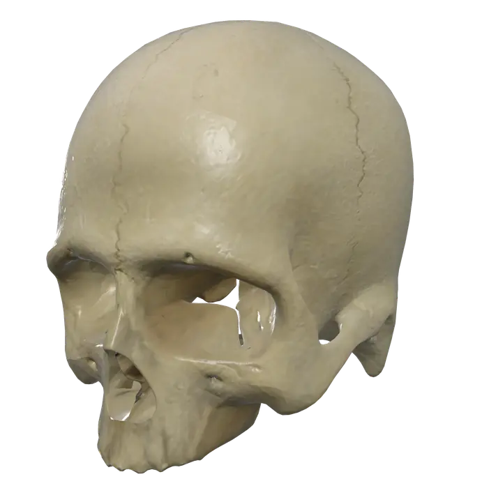
See it on the 3D body
The skull lit on the 3D head, as in the head quiz. Turn the body, peel the layers, tap any structure for its name.
Quick answer
- Latin name
- Cranium
- Type
- Group of flat and irregular bones joined by sutures
- Location
- Head, on top of the vertebral column
The adult skull is made of 22 bones: 8 cranial bones that enclose the brain and 14 facial bones that shape the face. All of them are locked together by fixed joints called sutures, except the lower jaw, the mandible, which moves at the temporomandibular joints.
The skull in Anatomy Atlas 3D
In the app, the skull is one structure, "Skull", covering the cranium and the upper face, while the mandible is a separate structure you can select on its own. That matches how the head works: the mandible is the only bone of the skull that moves.
This page describes the bones inside the "Skull" structure, so you know what to look for when you turn it in 3D.
Cranial bones
The cranium, or braincase, surrounds and protects the brain. Its eight bones are:
| Bone | Number | Where it is |
|---|---|---|
| Frontal | 1 | Forehead and the roof of the orbits |
| Parietal | 2 | Top and upper sides of the head |
| Temporal | 2 | Lower sides, around the ear |
| Occipital | 1 | Back and base of the skull |
| Sphenoid | 1 | Middle of the base, behind the eyes, shaped like a butterfly |
| Ethmoid | 1 | Between the orbits, behind the nose |
The temporal bone houses the middle and inner ear, with its small ear bones, the auditory ossicles. The sphenoid links almost every other cranial bone and holds the pituitary gland in a saddle-shaped hollow, the sella turcica. The ethmoid forms part of the walls of the nasal cavity and, with its perforated cribriform plate, part of the floor of the cranium.
Facial bones
The 14 facial bones form the face, the orbits, the nose and the mouth:
- Maxillae (2): the upper jaw, carrying the upper teeth and most of the hard palate.
- Zygomatic bones (2): the cheekbones.
- Nasal bones (2): the bridge of the nose.
- Lacrimal bones (2): tiny bones in the inner wall of each orbit.
- Palatine bones (2): the back of the hard palate.
- Inferior nasal conchae (2): curved plates on the side walls of the nasal cavity.
- Vomer (1): part of the nasal septum.
- Mandible (1): the lower jaw, separate in the app.
Sutures
Sutures are fibrous joints that allow almost no movement in adults. The main ones on the outside of the cranium:
- The coronal suture, across the top, between the frontal and the two parietal bones.
- The sagittal suture, along the midline, between the two parietal bones.
- The lambdoid suture, at the back, between the parietal bones and the occipital bone. Its name comes from the Greek letter lambda, which it resembles.
- The squamous suture, on each side, between the parietal and temporal bones.
At birth, the bones are not yet joined: soft membranous areas called fontanelles sit where several bones meet. They let the skull change shape during birth and grow with the brain. Most close during the first two years.
Key landmarks
Base and back of the skull
- The foramen magnum, a large opening in the occipital bone, where the brainstem continues as the spinal cord.
- The occipital condyles, on either side of it, which rest on the atlas at the atlanto-occipital joints.
- The external occipital protuberance, the bump you feel in the midline at the back of the head, and the superior nuchal lines running out from it.
Sides of the skull
- The mastoid process, the rounded bump behind the ear, part of the temporal bone.
- The styloid process, a thin spike below the ear.
- The external acoustic meatus, the bony opening of the ear canal.
- The zygomatic arch, the bar of bone along the cheek, formed by the temporal and zygomatic bones.
- The mandibular fossa, just in front of the ear, where the mandible articulates.
Front and inside
- The orbits, the sockets of the eyes, each built from seven bones.
- The nasal aperture, the opening of the nose in the dried skull.
- The anterior, middle and posterior cranial fossae, three steps in the floor of the cranium that hold the frontal lobes, the temporal lobes and the cerebellum.
- The paranasal sinuses, air spaces inside the frontal, maxillary, sphenoid and ethmoid bones.
Muscles that attach to the skull
- The temporalis starts on the side of the cranium, across the frontal, parietal, sphenoid and temporal bones, and closes the jaw.
- The masseter starts on the zygomatic arch and inserts on the mandible; both close the jaw.
- The sternocleidomastoid inserts on the mastoid process and the superior nuchal line.
- The trapezius starts on the external occipital protuberance and the superior nuchal line.
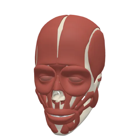
- Most muscles of facial expression start on the maxillae, zygomatic and nasal bones and insert into the skin. See the muscles of the face.
Frequently asked questions
How many bones are in the skull?
Usually 22 in the adult: 8 cranial and 14 facial bones. The six ear ossicles and the hyoid bone are generally counted separately.
Is the mandible part of the skull?
Yes, it is one of the 14 facial bones, and the only skull bone that moves. In Anatomy Atlas 3D it is a separate structure from the "Skull".
What are the 8 cranial bones?
The frontal, the two parietals, the two temporals, the occipital, the sphenoid and the ethmoid.
What is the foramen magnum?
The large opening in the occipital bone at the base of the skull, where the brainstem joins the spinal cord.
What are sutures in the skull?
Fibrous joints that hold the skull bones together and allow almost no movement in adults. The main ones are the coronal, sagittal, lambdoid and squamous sutures.
Muscles that originate here
- Trapezius
- Deep part of masseter
- Superficial part of masseter
- Temporalis
- Zygomaticus major
- Zygomaticus minor
- Buccinator
- Depressor septi nasi
- Levator anguli oris
- Levator labii superioris
- Levator labii superioris alaeque nasi
- Nasalis
- Procerus
Muscles that insert here
From the anatomical data behind Anatomy Atlas 3D (Z-Anatomy, reviewed). In the app, tap the bone, then each muscle around it to open its card.
