
Muscle · Head and neck
Posterior scalene: origin, insertion, action and innervation
The posterior scalene runs from C4-C6 to the second rib. It lifts that rib in deep breathing, bends the neck sideways and is supplied by cervical nerves.
Quick answer
- Latin name
- Musculus scalenus posterior
- Origin
- Posterior tubercles of the transverse processes of the fourth to sixth cervical vertebrae (C4–C6)
- Insertion
- Outer surface of the second rib
- Action
- Elevates the second rib in forced (accessory) inspiration. One side: bends the cervical spine laterally to the same side
- Innervation
- Anterior rami of the lower cervical spinal nerves (C6–C8; some textbooks give C7–C8)
- Blood supply
- Ascending cervical artery and branches of the transverse cervical artery
See it on the 3D body
The scalenus posterior lit on the 3D neck, as in the neck quiz. Turn the body, peel the layers, tap any structure for its name.
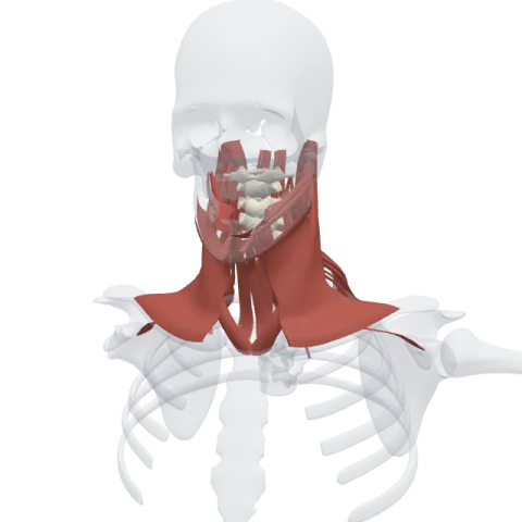
The posterior scalene is the smallest and deepest of the three scalene muscles. It runs from the lower cervical vertebrae to the second rib, which it lifts during deep breathing, and it bends the neck to the same side.
Where it is
The posterior scalene lies at the back of the scalene group, tucked behind the middle scalene and often partly fused with it. It sits low in the neck, just in front of the levator scapulae and under the upper fibers of the trapezius.
It is the only scalene that skips the first rib. Its fibers pass over the outside of the first rib and attach to the second, so it is easy to lose in a dissection: you have to follow the scalenes down past the first rib to find it.
Origin and insertion
- Origin: the posterior tubercles of the transverse processes of the cervical vertebrae C4 to C6.
- Insertion: the outer surface of the second rib, behind the area where the serratus anterior takes its origin.
Of the three scalenes, it has the shortest origin (three vertebrae) and the lowest insertion.
What it does
- Lifts the second rib: with the neck held steady, it raises the second rib. This adds a little to the expansion of the upper chest during forced inspiration, together with the anterior and middle scalenes lifting the first rib.
- Bends the neck to the side: acting on one side with the ribs held in place, it draws the lower cervical vertebrae down toward the second rib and tips the neck sideways.
Its effect on the ribs counts most when you need more air, for example during hard exercise.
Innervation and blood supply
The posterior scalene is supplied by small branches from the anterior rami of the lower cervical spinal nerves, C6 to C8 (some textbooks give only C7 and C8). The anterior and middle scalenes share the same type of supply from higher roots.
It is fed by the ascending cervical artery and by branches of the transverse cervical artery, the same small vessels of the lower neck that supply the middle scalene.
How to remember it
- P for passing: the posterior scalene passes over the first rib and lands on the second. Anterior and middle share rib one.
- Last, smallest, deepest: the posterior scalene is the runt of the group, smaller than the anterior scalene and far smaller than the middle one.
- Count the origins: anterior C3–C6, middle C2–C7, posterior C4–C6. The posterior scalene has the fewest starting points.
- Follow it over the first rib: if a muscle in the neck crosses the first rib without stopping, it is the posterior scalene.
Compare the first and second ribs on the bones of the rib cage page, then test yourself in the neck quiz.
Frequently asked questions
Which rib does the posterior scalene attach to?
The second rib, on its outer surface. The anterior and middle scalenes attach to the first rib.
What is the action of the posterior scalene?
It lifts the second rib in forced breathing and bends the cervical spine to the same side when one side contracts.
Which is the smallest scalene muscle?
The posterior scalene. It is also the deepest and the most posterior of the three.
What nerve supplies the posterior scalene?
Branches from the anterior rami of the lower cervical nerves, C6 to C8 (some textbooks say C7 and C8).
