
Muscle · Head and neck
Superior auricular muscle: origin, insertion, action and innervation
The superior auricular muscle fans from the epicranial aponeurosis to the top of the ear. It lifts the auricle and is supplied by the facial nerve (VII).
Quick answer
- Latin name
- Musculus auricularis superior
- Origin
- Epicranial aponeurosis (galea aponeurotica) at the side of the head, over the temporal fascia
- Insertion
- Upper part of the back (cranial surface) of the auricle
- Action
- Elevates the auricle
- Innervation
- Temporal branch of the facial nerve (VII)
- Blood supply
- Superficial temporal artery and posterior auricular artery
See it on the 3D body
The superior auricular lit on the 3D head, as in the head quiz. Turn the body, peel the layers, tap any structure for its name.
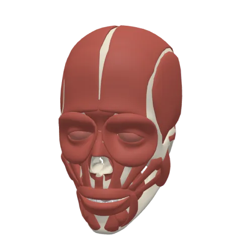
The superior auricular muscle is a thin, fan-shaped muscle on the side of the head that runs from the tendon sheet of the scalp down to the top of the ear. It lifts the auricle slightly, and it is supplied by the temporal branch of the facial nerve.
Where it is
The superior auricular muscle lies directly above the ear, on the side of the head, just under the skin and hair of the temple. It is the largest of the three extrinsic muscles of the ear, yet it is still very thin, a pale fan of fibers that is easy to miss.
Its fibers converge from above like a fan closing toward its handle: wide at the top, on the side of the scalp, and narrow at the bottom, where they reach the ear.
Under it lies the temporal fascia, the tough sheet that covers the temporalis, the big chewing muscle of the temple. The two muscles are stacked on top of each other but do completely different jobs: the temporalis closes the jaw, while the superior auricular muscle only moves the ear.
The other two ear muscles complete the set:
- the anterior auricular muscle, in front of the ear, which pulls it forward;
- the posterior auricular muscle, behind the ear, which pulls it backward.
Origin and insertion
- Origin: the epicranial aponeurosis, also called the galea aponeurotica, the flat sheet of tendon that covers the top of the skull and links the front and back bellies of the occipitofrontalis. At the side of the head this sheet thins out over the temporal fascia, and the superior auricular muscle arises from it there.
- Insertion: the upper part of the cranial surface of the auricle, the side of the ear that faces the head.
It has no attachment to bone at either end. It runs from a sheet of tendon to a piece of cartilage.
What it does
The superior auricular muscle elevates the auricle, pulling the ear upward. In humans the movement is small, and many people cannot make it on purpose. In animals that turn their ears toward sounds, muscles like this one are much stronger.
People who can move their ears may lift them with this muscle, often together with the scalp muscles that tighten the epicranial aponeurosis. Since the muscle starts on that sheet, raising the eyebrows or tensing the scalp can make the ear shift slightly too.
Like the other auricular muscles, it belongs to the muscles of facial expression: it moves skin and cartilage rather than a joint, and it shares their nerve, the facial nerve.
Innervation and blood supply
The superior auricular muscle is supplied by the temporal branch of the facial nerve (cranial nerve VII). The temporal branches leave the parotid gland, cross the zygomatic arch and run upward over the temple, where they supply the superior and anterior auricular muscles, the frontalis and the upper part of the orbicularis oculi.
Its blood comes mainly from the superficial temporal artery, which climbs in front of the ear, with a contribution from the posterior auricular artery behind the ear.
How to remember it
- Superior = above, so it lifts: each auricular muscle pulls the ear toward its own side.
- Biggest of three: superior is the largest auricular muscle, posterior and anterior are smaller.
- Temporal over temporalis: the temporal branch of the facial nerve supplies it, and it lies over the temporalis muscle.
- Tendon to cartilage: from the galea above to the ear below, no bone involved.
Place it among the other muscles of facial expression on the muscles of the face page, and read about the parts of the ear on the ear page. Test yourself in the head quiz.
Frequently asked questions
What does the superior auricular muscle do?
It elevates the auricle, pulling the ear upward. In most people the movement is very small.
Which nerve supplies the superior auricular muscle?
The temporal branch of the facial nerve (VII).
Which is the largest auricular muscle?
The superior auricular muscle. The anterior and posterior auricular muscles are smaller.
Where does the superior auricular muscle attach?
It arises from the epicranial aponeurosis on the side of the head and inserts on the upper part of the back of the auricle.
