Quick answer
- Latin name
- Larynx
- System
- Respiratory system
- Location
- Front of the neck, between the pharynx and the trachea
- Function
- Keeps food out of the airway, lets air pass and produces the voice
- Parts in the app
- 7
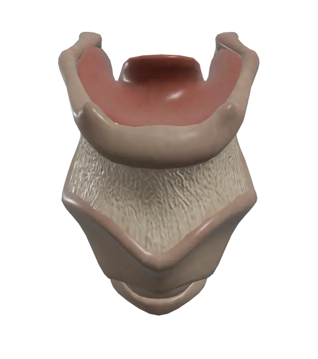
The larynx, often called the voice box, is a short tube of cartilage at the top of the airway. It connects the throat to the trachea and has three jobs: it lets air in and out, it stops food and drink from entering the lungs, and it holds the vocal folds that produce your voice.
Where it is
The larynx sits in the front of the neck, below the hyoid bone and above the trachea. Behind it is the lower part of the pharynx, which leads into the esophagus, so air and food share a crossroads right here. The thyroid gland wraps around the trachea just below the larynx, and the sternocleidomastoid muscles run diagonally on each side of the neck.
Its framework is made of cartilages joined by membranes and ligaments. Three are single: the thyroid cartilage, the cricoid cartilage below it (the only complete ring of cartilage in the airway) and the epiglottis. Three are paired and smaller: the arytenoid, corniculate and cuneiform cartilages, which sit at the back.
You can feel parts of the larynx with your fingers: the notch at the top of the thyroid cartilage, the soft gap below it, and the firm ring of the cricoid cartilage. When you swallow, the whole larynx rises.
Parts you can explore in the app
In the app you can open the larynx on its own and study it part by part:
- Thyroid cartilage: the largest cartilage of the larynx, made of two plates that meet in front to form the laryngeal prominence, or Adam's apple.
- Thyrohyoid membrane: a sheet of connective tissue that links the upper edge of the thyroid cartilage to the hyoid bone above.
- Cricothyroid ligament: connects the cricoid cartilage to the thyroid cartilage. Its middle part lies just under the skin, below the Adam's apple.
- Epiglottis: a leaf-shaped flap of elastic cartilage behind the root of the tongue that tips over the entrance to the larynx when you swallow.
- Epiglottis (inner surface): the side of the epiglottis that faces into the airway, covered by a mucous membrane.
- Vocal fold (left) and Vocal fold (right): the true vocal cords, folds of mucous membrane over a ligament and muscle, stretched from the thyroid cartilage in front to the arytenoid cartilages behind. The gap between them is the glottis.
Cross sections (sagittal, coronal and axial) let you look down into the larynx and see how narrow the passage becomes at the level of the vocal folds.
What it does
Breathing. When you breathe in, the vocal folds move apart so air can pass freely between the throat and the trachea.
Protecting the airway. During swallowing, the larynx is pulled upward and forward, the epiglottis folds back over the laryngeal inlet, and the vocal folds close. Food and liquid slide past into the esophagus instead of the airway. If something does get in, sensitive nerve endings in the lining trigger a cough.
Making sound. When the vocal folds come together and air from the lungs is pushed between them, they vibrate and produce sound. Pitch depends on how tense and long the folds are; tighter folds vibrate faster and give a higher note. Loudness depends mostly on how hard the air is pushed. The throat, mouth, tongue and lips then shape this raw sound into speech.
Most of the small muscles inside the larynx are supplied by the recurrent laryngeal nerve, a branch of the vagus nerve. The left recurrent laryngeal nerve loops under the arch of the aorta and the right one under the right subclavian artery before climbing back up to the larynx. The cricothyroid muscle, which tightens the vocal folds, is supplied by the superior laryngeal nerve instead.
How to study it
- Three single, three paired. Thyroid, cricoid and epiglottis are single; arytenoid, corniculate and cuneiform are paired.
- Feel it on yourself. Run a finger down the front of your neck: hyoid bone, thyroid notch, the soft gap of the cricothyroid ligament, then the cricoid ring. Linking each structure to a spot you can touch makes it stick.
- True versus false. The vocal folds (true cords) produce the voice; the vestibular folds (false cords) lie just above them and mainly play a protective role.
- The nerve exception. Every intrinsic muscle of the larynx is supplied by the recurrent laryngeal nerve except the cricothyroid, which is supplied by the superior laryngeal nerve. Exams love this.
- Use the coronal section to see the vocal folds and the vestibular folds as two pairs of ridges on the inner walls.
Frequently asked questions
What is the Adam's apple?
It is the laryngeal prominence, the point where the two plates of the thyroid cartilage meet at the front of the neck. It usually becomes more visible in males after puberty.
What does the epiglottis do?
It acts like a lid. During swallowing it folds back over the entrance to the larynx and helps guide food and liquid into the esophagus.
How do the vocal folds make sound?
Air pushed up from the lungs makes the closed vocal folds vibrate. Changing their tension changes the pitch, and changing the air pressure changes the loudness.
Is the larynx part of the respiratory system?
Yes. It is part of the upper airway, between the pharynx and the trachea, and it also plays a key role in swallowing and speech.