
Muscle · Upper limb
Dorsal interossei of the hand: origin, insertion, action and innervation
The four dorsal interossei of the hand lie between the metacarpals. They spread the fingers apart and are supplied by the deep branch of the ulnar nerve.
Quick answer
- Latin name
- Musculi interossei dorsales manus
- Origin
- Adjacent sides of the five metacarpals: each of the four muscles has two heads, one from each bone that borders its space
- Insertion
- Bases of the proximal phalanges and extensor expansions: radial side of the index and middle fingers, ulnar side of the middle and ring fingers
- Action
- Abduct the index, middle and ring fingers at the metacarpophalangeal joints, away from the axis of the middle finger; help flex the knuckles and extend the finger joints
- Innervation
- Deep branch of the ulnar nerve (C8-T1)
- Blood supply
- Dorsal and palmar metacarpal arteries
See it on the 3D body
The dorsal interossei muscles of hand lit on the 3D forearm and hand, as in the forearm and hand quiz. Turn the body, peel the layers, tap any structure for its name.
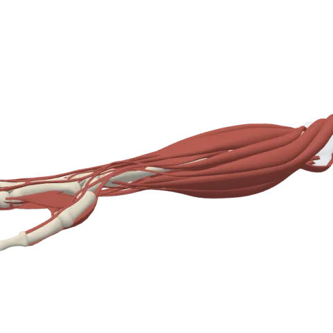
The dorsal interossei of the hand are four small muscles that fill the spaces between the metacarpal bones and spread the fingers apart. They abduct the index, middle and ring fingers away from the middle finger and are supplied by the deep branch of the ulnar nerve.
Where it is
Interossei means "between the bones". The dorsal interossei sit in the four gaps between the five metacarpals, closer to the back of the hand than the palmar interossei, which share the same spaces on the palm side. They are numbered from the thumb side: the first lies between the first and second metacarpals, the fourth between the fourth and fifth.
Each dorsal interosseous is bipennate: it has two heads, one from each bone that borders its space, and the fibers meet on a central tendon like the vanes of a feather.
The first dorsal interosseous is the largest. It fills the web between the thumb and the index finger on the back of the hand. Squeeze your index finger against the thumb and you can see it bulge there. The radial artery passes between its two heads to enter the palm.
Origin and insertion
- Origin: the facing sides of two neighboring metacarpals, for each of the four muscles.
- Insertion: the base of the proximal phalanx and the extensor expansion of a finger, on the side that points away from the middle finger:
- first: radial (thumb) side of the index finger;
- second: radial side of the middle finger;
- third: ulnar (little-finger) side of the middle finger;
- fourth: ulnar side of the ring finger.
The middle finger receives two dorsal interossei, one on each side. The thumb and the little finger receive none, because they have their own abductors, the abductor pollicis brevis and the abductor digiti minimi. See the phalanges of the hand for the insertion points.
What it does
- Abduction of the fingers: at the metacarpophalangeal joints, they move the index, middle and ring fingers away from an imaginary line drawn through the middle finger. This line, not the midline of the body, is the reference for finger movements.
- Side-to-side movement of the middle finger: with a muscle on each side, the middle finger can be pulled toward the thumb or toward the little finger. Both movements count as abduction.
- Flexion of the knuckles with straight fingers: because they pass in front of the axis of the knuckle joints and then join the extensor expansion, they help bend the knuckles while keeping the finger joints straight, the position of a hand holding a tray. The lumbricals and the palmar interossei share this job, and the extensor digitorum relies on them for full finger extension.
Innervation and blood supply
All four are supplied by the deep branch of the ulnar nerve (roots C8 and T1). The branch crosses the palm and sends a twig to each interosseous muscle on its way to the thumb. When the ulnar nerve is damaged, the first dorsal interosseous can waste away and leave a visible hollow in the web on the back of the hand.
Their blood comes from the dorsal metacarpal arteries on the back of the hand and the palmar metacarpal arteries, branches of the deep palmar arch.
How to remember it
- DAB and PAD: Dorsal interossei ABduct, Palmar interossei ADduct.
- Count from the middle finger: abduction of the fingers means moving away from the middle finger, so the middle finger needs a muscle on each side.
- Four spaces, four muscles: one dorsal interosseous per gap between the metacarpals, numbered from the thumb.
- Thumb and pinky bring their own: they have private abductors, so they need no dorsal interosseous.
Test yourself in the forearm and hand quiz, and find the metacarpals on the bones of the hand page.
Frequently asked questions
What do the dorsal interossei of the hand do?
They abduct the index, middle and ring fingers at the metacarpophalangeal joints, away from the middle finger. They also help flex the knuckles and extend the finger joints.
How many dorsal interossei are there in the hand?
Four, one in each space between the five metacarpals.
Which nerve supplies the dorsal interossei?
All four get the deep branch of the ulnar nerve (C8-T1).
What is the difference between dorsal and palmar interossei?
The dorsal interossei spread the fingers apart (abduction) and have two heads each. The palmar interossei bring the fingers together (adduction) and have one head each.
Why does the middle finger have two dorsal interossei?
Because finger movements are measured from the middle finger. It can move to either side, and each movement is abduction, so it needs one muscle on each side.
