
See them on the 3D body
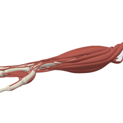
The phalanges of the hand lit on the 3D forearm and hand, as in the forearm and hand quiz. Turn the body, peel the layers, tap any structure for its name.
Quick answer
- Latin name
- Ossa digitorum manus (phalanges)
- Type
- Long bones (fourteen phalanges per hand)
- Location
- Fingers and thumb, beyond the heads of the metacarpals
- Joints
- Metacarpophalangeal joints with the metacarpals; proximal and distal interphalangeal joints in the fingers; a single interphalangeal joint in the thumb
The phalanges of the hand are the bones of the fingers and thumb. Each finger has three, called the proximal, middle and distal phalanx, while the thumb has only two, a proximal and a distal one. That makes 14 phalanges in each hand.
Where they are and what they touch
The phalanges start beyond the knuckles. The proximal phalanx of each digit joins the head of its metacarpal, the middle phalanx follows, and the distal phalanx forms the tip of the finger under the nail. The fingers are numbered from the thumb: thumb (1), index (2), middle (3), ring (4) and little finger (5).
The phalanges get shorter from base to tip, and the middle finger has the longest ones. They carry no muscle bellies of their own: they are moved by tendons that come from the forearm and from the small muscles of the palm. The bones of the hand page shows how they fit with the carpals and metacarpals.
Parts of a phalanx
Every phalanx is a miniature long bone with three parts:
- a base at the near end, with a joint surface;
- a shaft, flat on the palm side and rounded on the back;
- a head at the far end.
The shape of the ends changes from one row to the next, and that shape decides how each joint moves.
Proximal phalanges
The proximal phalanges are the longest of each digit. Their base has an oval, hollow surface that fits on the rounded head of the metacarpal. Their head is shaped like a pulley, with two small rounded knobs and a groove between them, which fits the base of the middle phalanx.
The dorsal interossei insert partly on their bases, and the interossei and lumbricals also join the extensor expansion that covers their backs. The small muscles of the little finger insert on the base of its proximal phalanx.
Middle phalanges
Only the four fingers have a middle phalanx. Its base has two shallow hollows separated by a ridge, matching the pulley of the proximal phalanx. Its head is again pulley-shaped. The two slips of the flexor digitorum superficialis tendon insert on the sides of its shaft, and the central band of the extensor tendon reaches the back of its base.
Distal phalanges
The distal phalanges are the smallest. Their tip widens into a rough, horseshoe-shaped tuberosity that supports the pad of the fingertip and the nail bed. The flexor digitorum profundus tendon inserts on the palm side of their base, and the end of the extensor tendon on the back.
The thumb: two phalanges
The thumb has a proximal and a distal phalanx, but no middle one. Both are shorter and wider than those of the fingers.
- The proximal phalanx of the thumb receives the extensor pollicis brevis on its back and the adductor pollicis on its inner side, along with two short thumb muscles on its outer side.
- The distal phalanx of the thumb receives the flexor pollicis longus on its palm side and the extensor pollicis longus on its back.
Two small sesamoid bones usually sit in tendons on the palm side of the thumb's knuckle joint.
Joints
- Metacarpophalangeal (MCP) joints: between the metacarpal heads and the proximal phalanges. The fingers can bend, straighten, spread and close here. The thumb's MCP joint moves mostly as a hinge.
- Proximal interphalangeal (PIP) joints: between the proximal and middle phalanges of the fingers.
- Distal interphalangeal (DIP) joints: between the middle and distal phalanges.
- Interphalangeal (IP) joint of the thumb: the only joint between its two phalanges.
The PIP, DIP and thumb IP joints are hinge joints: they only bend and straighten, held on each side by collateral ligaments.
Muscles attached
The long tendons come from the muscles of the forearm:
- The extensor digitorum sends a tendon to each finger, which spreads into the extensor expansion and inserts on the middle and distal phalanges. It straightens the fingers.
- The extensor indicis and the extensor digiti minimi join this expansion for the index and little fingers.
- The flexor digitorum superficialis inserts on the middle phalanges and bends the PIP joints.
- The flexor digitorum profundus inserts on the distal phalanges and is the only muscle that bends the DIP joints.
- In the thumb, the long flexor and long extensor insert on the distal phalanx.
How to remember them
- Three, three, three, three, two. Count the phalanges from little finger to thumb: 3 + 3 + 3 + 3 + 2 = 14.
- Superficial splits, deep goes deep. The superficialis tendon splits to let the profundus tendon through, and stops at the middle phalanx. The profundus continues to the distal phalanx.
- Phalanx, phalanges. One finger bone is a phalanx. The name comes from a row of soldiers, which is how they line up.
- Short and wide means thumb. If a phalanx is stubby, it is probably from the thumb.
In Anatomy Atlas 3D
The app names every phalanx by row and finger, for example "Middle phalanx of ring finger" or "Distal phalanx of thumb". Turn the hand palm up and tap each tip to check you can tell the fingers apart. Practice them in the forearm and hand quiz. Anatomy Atlas 3D is free on iPad and iPhone.
Frequently asked questions
Why does the thumb have only two phalanges?
It has a proximal and a distal phalanx but no middle one. Its range of movement comes instead from its metacarpal and the saddle joint at the wrist.
Which muscle bends the fingertip?
The flexor digitorum profundus. Its tendons insert on the base of each distal phalanx of the four fingers.
What is the tuberosity of the distal phalanx?
The widened, rough tip of each distal phalanx. It supports the fingertip pad and the nail bed.
What are the PIP and DIP joints?
The proximal and distal interphalangeal joints of the fingers. The PIP joint is between the proximal and middle phalanges, the DIP joint between the middle and distal phalanges.
From the anatomical data behind Anatomy Atlas 3D (Z-Anatomy, reviewed). In the app, tap the bone, then each muscle around it to open its card.
