
Muscle · Head and neck
Orbicularis oculi: origin, insertion, action and innervation
The orbicularis oculi rings the eye, from the medial orbital margin to the skin around the orbit. It closes the eyelids and is supplied by the facial nerve.
Quick answer
- Latin name
- Musculus orbicularis oculi
- Origin
- Medial margin of the orbit (nasal part of the frontal bone, frontal process of the maxilla, lacrimal bone) and the medial palpebral ligament
- Insertion
- Skin around the orbit and the lateral palpebral raphe
- Action
- Closes the eyelids: gently for blinking and sleep (palpebral part), tightly for squeezing (orbital part)
- Innervation
- Temporal and zygomatic branches of the facial nerve (VII)
- Blood supply
- Branches of the facial, superficial temporal and ophthalmic arteries
See it on the 3D body
The orbicularis oculi lit on the 3D head, as in the head quiz. Turn the body, peel the layers, tap any structure for its name.
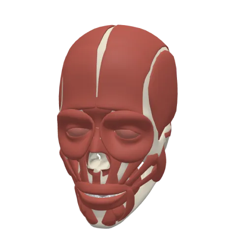
The orbicularis oculi is the flat ring of muscle that surrounds the eye and fills the eyelids. It closes the eyelids, both for a quick blink and for squeezing the eyes tightly shut, and it is supplied by the temporal and zygomatic branches of the facial nerve.
Where it is
The orbicularis oculi lies just under the thin skin around the eye. It covers the eyelids and spreads beyond them over the bony rim of the orbit, onto the forehead, the temple and the upper cheek. Its fibers form loops around the opening between the eyelids, like the rings of a target.
It has three parts:
- The palpebral part, thin and pale, inside the upper and lower eyelids.
- The orbital part, thicker and redder, on the bony margin of the orbit around the eyelids.
- The lacrimal part (deep part), small, behind the lacrimal sac on the inner side of the orbit.
Above it lie the eyebrow and the frontalis; at its inner upper corner it meets the procerus and the corrugator supercilii. The eyeball itself sits behind it, inside the bony orbit of the skull. See the eye page for the structures behind the lids.
Origin and insertion
The ring is anchored mainly on the inner side of the orbit:
- Origin: the medial margin of the orbit, made of the nasal part of the frontal bone and the frontal process of the maxilla; the medial palpebral ligament, a short band at the inner corner of the eye; and, for the lacrimal part, the lacrimal bone behind the lacrimal sac.
- Insertion: the fibers loop around the eye. Palpebral fibers meet at the outer corner in a seam called the lateral palpebral raphe. Orbital fibers make complete loops and blend with the skin and the neighboring muscles.
What it does
- Gentle closure: the palpebral part closes the eyelids without effort, in blinking and in sleep. Blinking spreads the tear film over the front of the eye and keeps it moist.
- Tight closure: the orbital part squeezes the eyes shut, pulling the skin of the forehead, temple and cheek toward the inner corner. This protects the eye from glare, wind or a blow, and it creases the skin at the outer corner.
- Tear drainage: the lacrimal part pulls the eyelids against the eyeball and acts on the lacrimal sac, which helps move tears toward the nose.
Its opponent is the levator palpebrae superioris, the muscle that lifts the upper eyelid, supplied by the oculomotor nerve. In a genuine smile, the orbital part joins the zygomaticus major and narrows the eyes.
Innervation and blood supply
The orbicularis oculi is supplied by the temporal and zygomatic branches of the facial nerve (cranial nerve VII). The temporal branches reach the upper part of the ring, the zygomatic branches the lower part. In facial nerve palsy, the eyelids on the affected side cannot close fully.
Its blood comes from several small arteries around the eye: branches of the facial artery (angular artery), of the superficial temporal artery (zygomatico-orbital and transverse facial arteries) and of the ophthalmic artery (palpebral, supraorbital and lacrimal arteries).
How to remember it
- ***Orbis* = circle, oculus = eye**: the circle around the eye.
- Palpebral for blinking, orbital for squeezing: the thin inner ring does the daily work, the thick outer ring the hard squeeze.
- Closes, never opens: lifting the upper eyelid is the job of another muscle with another nerve (oculomotor, cranial nerve III).
- Temporal up, zygomatic down: two facial nerve branches split the ring between them.
The muscles of the face page shows how it works with the smile muscles. Test yourself in the head quiz.
Frequently asked questions
What is the function of the orbicularis oculi?
It closes the eyelids. Its palpebral part blinks and closes the eyes in sleep; its orbital part squeezes them tightly shut.
Which nerve supplies the orbicularis oculi?
The temporal and zygomatic branches of the facial nerve (VII).
What are the parts of the orbicularis oculi?
The palpebral part in the eyelids, the orbital part around the rim of the orbit and the small lacrimal part behind the lacrimal sac.
Which muscle opens the eye?
The levator palpebrae superioris, which lifts the upper eyelid. The orbicularis oculi only closes it.
