Quick answer
- Latin name
- Bulbus oculi
- System
- Nervous system (special senses)
- Location
- In the orbit of the skull, cushioned by fat
- Function
- Focuses light on the retina and turns it into nerve signals for vision
- Parts in the app
- 17
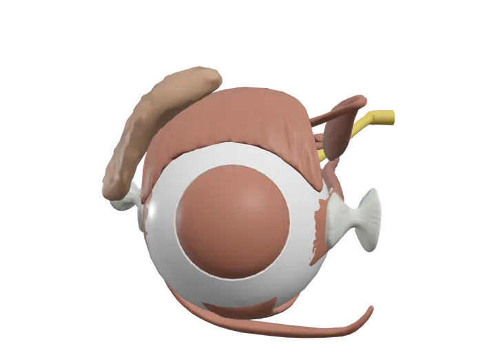
The eye is the organ of vision. It works a little like a camera: clear structures at the front bend light and focus it onto a light-sensitive layer at the back, which turns it into nerve signals for the brain. Six small muscles aim it, and the lacrimal gland keeps its surface moist.
Where it is
Each eyeball sits in the orbit, a bony socket of the skull, cushioned by fat. Only the front of the eye is exposed, protected by the eyelids and covered by a thin membrane, the conjunctiva. The extraocular muscles run from the back of the orbit to the eyeball, and the optic nerve leaves the back of the eye through the optic canal to reach the brain.
The wall of the eyeball has three layers:
- The outer fibrous layer: the sclera and the cornea.
- The middle vascular layer: the choroid, the ciliary body and the iris.
- The inner neural layer: the retina.
Parts you can explore in the app
In the app you can open the eye on its own and study it part by part:
- Sclera: the tough white outer coat. It gives the eyeball its shape and anchors the eye muscles.
- Cornea: the clear dome at the front. It does most of the work of bending light.
- Iris: the colored ring around the pupil. Its muscles make the pupil smaller or larger.
- Lens: a clear, flexible disc behind the iris that fine-tunes focus by changing shape.
- Retina: the inner light-sensitive layer, with rods and cones that respond to light.
- Vitreous body: the clear gel that fills the space behind the lens and helps keep the eye's shape.
- Anterior chamber: the fluid-filled space between the cornea and the iris, filled with aqueous humor.
- Suspensory ligament of eyeball: a sling of connective tissue under the eyeball that helps hold it in position in the orbit.
- Optic nerve: cranial nerve II, which carries visual signals from the retina to the brain.
- Superior rectus: lifts the gaze upward.
- Inferior rectus: lowers the gaze.
- Medial rectus: turns the eye toward the nose.
- Lateral rectus: turns the eye outward, away from the nose.
- Superior oblique: its tendon passes through a small pulley, the trochlea. It lowers the gaze, especially when the eye looks toward the nose, and rotates the eye inward.
- Inferior oblique: lifts the gaze, especially when the eye looks toward the nose, and rotates the eye outward.
- Levator palpebrae: raises the upper eyelid.
- Lacrimal gland: sits in the upper outer corner of the orbit and makes tears.
Cross sections (sagittal, coronal and axial) let you cut through the eyeball and see the chambers, the lens and the layers in order.
What it does
Light enters through the cornea, crosses the aqueous humor of the anterior chamber, passes through the pupil, then through the lens and the vitreous body before it reaches the retina. The cornea provides most of the focusing power; the lens adjusts it. To look at something close, the ciliary muscle contracts, the fibers holding the lens slacken, and the lens becomes rounder. This is called accommodation.
The iris controls how much light gets in. In bright light its circular muscle narrows the pupil; in dim light its radial muscle widens it.
The retina contains two kinds of photoreceptors. Rods work in dim light and give black-and-white vision. Cones need brighter light and give color and sharp detail. Cones are packed into the fovea, a small pit in the center of the macula, where vision is sharpest. Where the optic nerve leaves the eye, the optic disc has no photoreceptors at all, which creates a blind spot.
Signals travel along the optic nerve. At the optic chiasm, fibers from the nasal half of each retina cross to the other side, so each side of the brain sees the opposite half of the visual field. The pathway relays in the thalamus and ends in the occipital lobe.
The six extraocular muscles move both eyes together so their images line up. Tears from the lacrimal gland wash across the eye with each blink and drain through small openings near the inner corner of the eyelids into the nose.
How to study it
- Learn the three layers first. Fibrous, vascular, neural: almost every structure of the eyeball belongs to one of them.
- "LR6 SO4, the rest 3." The lateral rectus is supplied by the abducens nerve (VI), the superior oblique by the trochlear nerve (IV), and the other eye muscles, including levator palpebrae, by the oculomotor nerve (III).
- Trace the path of light. Cornea, aqueous humor, pupil, lens, vitreous body, retina. Exams often ask for this order.
- Rods for dark, cones for color. And remember that the fovea has only cones, while the optic disc has none.
- Watch the obliques. They attach behind the middle of the eyeball, so they act the "opposite" way to their names: the superior oblique lowers the gaze, the inferior oblique raises it.
- Use the sagittal section to see the chambers, lens and vitreous body in a single view.
Frequently asked questions
What are the three layers of the eye?
The fibrous layer (sclera and cornea), the vascular layer (choroid, ciliary body and iris) and the neural layer (retina).
Which part of the eye focuses light?
The cornea does most of the focusing. The lens then adjusts the focus for near or far objects by changing shape.
What are the six extraocular muscles?
The superior, inferior, medial and lateral rectus muscles, and the superior and inferior oblique muscles.
Why do we have a blind spot?
The optic disc, where the optic nerve leaves the eye, has no rods or cones. Light that lands there is not detected, but the brain fills in the gap.