
Muscle · Head and neck
Mylohyoid muscle: origin, insertion, action and innervation
The mylohyoid runs from the mylohyoid line of the mandible to the hyoid. It raises the floor of the mouth and is supplied by the nerve to mylohyoid.
Quick answer
- Latin name
- Musculus mylohyoideus
- Origin
- Mylohyoid line on the inner surface of the mandible
- Insertion
- Body of the hyoid bone and a midline fibrous seam (raphe) running from the chin to the hyoid
- Action
- Elevates the floor of the mouth and the hyoid bone during swallowing and speech; helps depress the mandible when the hyoid is fixed
- Innervation
- Nerve to mylohyoid, a branch of the inferior alveolar nerve (mandibular nerve, CN V3)
- Blood supply
- Mylohyoid branch of the inferior alveolar artery and the submental artery
See it on the 3D body
The mylohyoid lit on the 3D neck, as in the neck quiz. Turn the body, peel the layers, tap any structure for its name.
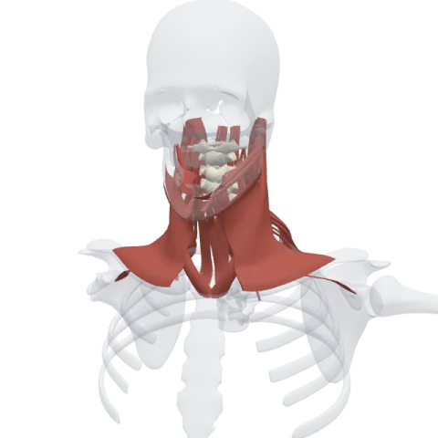
The mylohyoid is a flat, thin muscle that stretches across the inside of the lower jaw like a hammock and forms the muscular floor of the mouth. It lifts that floor, the tongue and the hyoid bone when you swallow and speak.
Where it is
The left and right mylohyoids meet in the midline under the tongue, so together they make one continuous sheet between the two halves of the mandible. Anatomists sometimes call this sheet the oral diaphragm.
It is one of the suprahyoid muscles. Its relations are worth learning layer by layer:
- Below it: the anterior belly of the digastric, the skin and the platysma.
- Above it: the geniohyoid muscle, the tongue and the floor of the mouth, including the sublingual salivary gland.
- Around its back edge: the submandibular salivary gland curls around the free posterior border of the muscle, with one part below it and one part above it.
The hyoglossus lies deep to the mylohyoid, against the side of the tongue.
Origin and insertion
- Origin: the mylohyoid line, a ridge that runs diagonally along the inner surface of the body of the mandible, from near the last molar toward the chin.
- Insertion: the front fibers meet their partners from the other side at a midline raphe, a fibrous seam from the chin down to the hyoid. The back fibers attach directly to the body of the hyoid bone.
The name tells you both ends: mylo comes from the Greek for "molar", referring to the molar region of the jaw, and hyoid refers to the hyoid bone.
What it does
- Raises the floor of the mouth: at the start of swallowing, it tightens and lifts the floor, pushing the tongue up against the palate so the food bolus moves back toward the throat.
- Lifts the hyoid bone: with the jaw closed, it pulls the hyoid up and forward, which helps raise the larynx during swallowing.
- Helps open the mouth: when the infrahyoid muscles hold the hyoid still, it can assist in pulling the mandible down.
It also adjusts the floor of the mouth during speech, giving the tongue a firm base to work against.
Innervation and blood supply
The mylohyoid is supplied by the nerve to mylohyoid. This small nerve branches from the inferior alveolar nerve just before that nerve enters the mandibular canal, so it belongs to the mandibular division of the trigeminal nerve (CN V3). It runs in a groove on the inner surface of the mandible and also supplies the anterior belly of the digastric.
Blood comes from the mylohyoid branch of the inferior alveolar artery, which travels with the nerve, and from the submental artery, a branch of the facial artery.
How to remember it
- Molar to hyoid: "mylo" sounds like "molar". The muscle starts near the molars and ends on the hyoid.
- A nerve named after its muscle: the nerve to mylohyoid supplies the mylohyoid, plus one extra: the anterior digastric just below it.
- Hammock under the tongue: picture two sheets of muscle hung from each side of the jaw, sewn together in the middle.
- Gland wrapped round the edge: the submandibular gland hooks around the back border of the mylohyoid like a folded towel over a rail.
The larynx page shows the structures the mylohyoid helps lift. Test yourself in the neck quiz.
Frequently asked questions
What does the mylohyoid muscle do?
It raises the floor of the mouth and the hyoid bone during swallowing and speech. It can also help open the mouth when the hyoid is fixed.
What nerve supplies the mylohyoid?
The nerve to mylohyoid, a branch of the inferior alveolar nerve from the mandibular nerve (CN V3).
Where is the mylohyoid line?
On the inner surface of the mandible. It is a diagonal ridge that runs from the molar area toward the chin.
Is the mylohyoid the floor of the mouth?
Yes. The two mylohyoid muscles, joined at a midline seam, form the muscular floor of the mouth.
