
Muscle · Head and neck
Stylohyoid muscle: origin, insertion, action and innervation
The stylohyoid runs from the styloid process to the hyoid bone. It pulls the hyoid up and back when you swallow and is supplied by the facial nerve.
Quick answer
- Latin name
- Musculus stylohyoideus
- Origin
- Back of the styloid process of the temporal bone (skull)
- Insertion
- Body of the hyoid bone, where it joins the greater horn
- Action
- Elevates and retracts the hyoid bone, lengthening the floor of the mouth during swallowing
- Innervation
- Stylohyoid branch of the facial nerve (CN VII)
- Blood supply
- Branches of the facial, posterior auricular and occipital arteries
See it on the 3D body
The stylohyoid lit on the 3D neck, as in the neck quiz. Turn the body, peel the layers, tap any structure for its name.
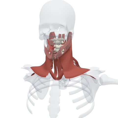
The stylohyoid is a thin muscle that runs from the styloid process, a needle of bone under the ear, down to the hyoid bone. It pulls the hyoid up and backward when you swallow, and it is supplied by the facial nerve.
Where it is
The stylohyoid lies in the upper neck, under the angle of the mandible. It belongs to the suprahyoid muscles, with the digastric, the mylohyoid and the geniohyoid.
It travels right alongside the posterior belly of the digastric, just above and in front of it. The two are so close that they are often studied as a pair. Near the hyoid, the stylohyoid splits into two slips, and the intermediate tendon of the digastric passes through the gap before the slips rejoin and attach to the bone.
The styloid process gives its name to three muscles: the stylohyoid, the styloglossus (to the tongue) and the stylopharyngeus (to the pharynx). The stylohyoid is the most superficial of the three.
Origin and insertion
- Origin: the back and side of the styloid process of the temporal bone, a slender spike that points down and forward from the base of the skull, just in front of the mastoid process.
- Insertion: the body of the hyoid bone, at the point where the body meets the greater horn.
The muscle is short and spindle-shaped, with a narrow tendon at its upper end.
What it does
- Pulls the hyoid up and back: when it contracts, the hyoid moves toward the styloid process, so it rises and slides backward.
- Lengthens the floor of the mouth: by drawing the hyoid back while the mylohyoid lifts the floor, it stretches the floor of the mouth front to back. This helps the tongue push food toward the throat during swallowing.
The stylohyoid works with the posterior belly of the digastric, which pulls in almost the same direction.
Innervation and blood supply
The stylohyoid is supplied by the stylohyoid branch of the facial nerve (CN VII). The branch leaves the facial nerve right after it exits the skull through the stylomastoid foramen, close to the branch for the posterior belly of the digastric. Both muscles come from the second pharyngeal arch, the arch served by the facial nerve.
Blood comes from small branches of the facial, posterior auricular and occipital arteries, all from the external carotid system.
How to remember it
- Read the name as a route: stylo-hyoid goes from the styloid process to the hyoid.
- Seven for the styloid twins: the stylohyoid and the posterior digastric run side by side and share cranial nerve VII.
- The muscle with a buttonhole: it is the one that lets the digastric tendon pass through it.
- Three stylo muscles, three nerves: stylohyoid by VII, stylopharyngeus by IX, styloglossus by XII.
Find the styloid process on the skull page, then test yourself in the neck quiz.
Frequently asked questions
What is the action of the stylohyoid muscle?
It elevates and retracts the hyoid bone. This lengthens the floor of the mouth during swallowing.
What nerve supplies the stylohyoid?
The stylohyoid branch of the facial nerve (CN VII).
Where does the stylohyoid originate?
From the styloid process of the temporal bone, under the ear.
How is the stylohyoid related to the digastric?
It runs alongside the posterior belly of the digastric, and near the hyoid it splits around the digastric's intermediate tendon.
