
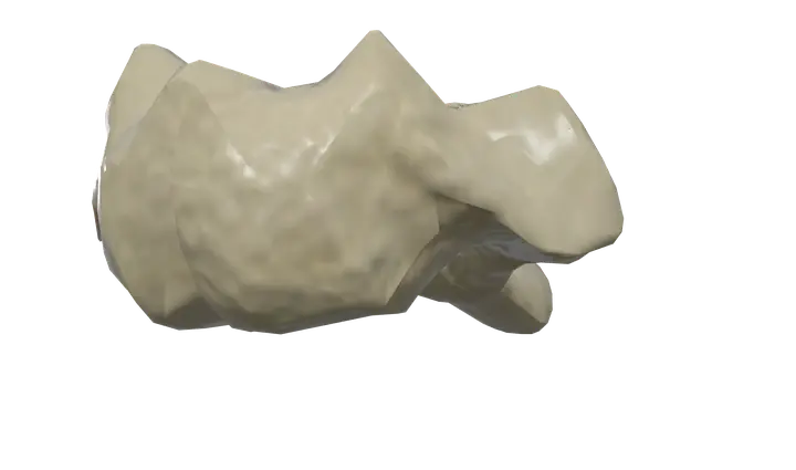
See it on the 3D body
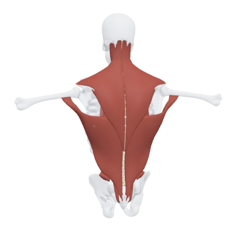
The axis (C2 vertebra) lit on the 3D back, as in the back quiz. Turn the body, peel the layers, tap any structure for its name.
Quick answer
- Latin name
- Axis
- Type
- Irregular bone (atypical cervical vertebra)
- Location
- Upper neck, just below the atlas
- Joints
- Atlanto-axial joints with the atlas; intervertebral disc and facet joints with the third cervical vertebra (C3)
The axis is the second cervical vertebra (C2), recognizable by the dens, a tooth-like peg of bone that rises from its body into the ring of the atlas. The atlas, carrying the skull, turns around this peg, which is why the axis is where most of the head's side-to-side rotation happens.
Where it is and what it touches
The axis sits just below the atlas, at the top of the neck. Its large spinous process is the first bony bump you meet in the midline when you run a finger down from the base of the skull.
It has two kinds of joints:
- Above, with the atlas, at the atlanto-axial joints. In the middle, the dens fits against the back of the anterior arch of the atlas, held there by the transverse ligament: this is the median atlanto-axial joint, a pivot joint. On each side, the broad superior facets of the axis meet the lateral masses of the atlas at the lateral atlanto-axial joints.
- Below, with the third cervical vertebra (C3), like a typical vertebra: an intervertebral disc between the bodies, the first disc of the spine, and two facet joints behind.
The bones of the spine page shows the axis in the cervical spine with the other vertebrae.
Shape and landmarks
The axis combines an unusual top with a fairly typical bottom.
Dens
- Also called the odontoid process, from the Greek for tooth.
- A short vertical peg rising from the top of the body.
- Its front has an anterior articular facet for the anterior arch of the atlas; its back has a smooth groove, the posterior articular facet, for the transverse ligament.
- Its tip anchors small ligaments that tie it to the base of the skull: the apical ligament at the very tip and the two alar ligaments, which run from its sides to the edges of the foramen magnum and limit how far the head can turn.
Body
- The body of the axis lies below the dens. Developmentally, the dens is the body of the atlas, fused onto the axis.
- Its lower edge projects downward in front, overlapping the top of C3.
Articular facets
- The superior articular facets are large, flat and oval, and sit on either side of the dens, more forward than the facets of the vertebrae below. They carry the weight of the head and atlas.
- The inferior articular facets lie farther back, in line with those of the lower cervical vertebrae, and face down and forward toward C3.
Arch and processes
- Pedicles and laminae form the arch around the spinal cord. The laminae are thick and strong.
- The transverse processes are small and each has a transverse foramen for the vertebral artery.
- The spinous process is large and bifid, split into two tips at its end. It is the biggest spinous process of the upper neck and anchors several deep neck muscles.
How the atlas and axis work together
The atlas and axis are built as a pair. The atlas has no body, and its missing body forms the dens of the axis. The atlas is a ring; the axis provides the pivot.
- Rotation. When you shake your head "no", the atlas and skull turn together around the dens. Roughly half of the rotation of the whole neck takes place here.
- Nodding. When you nod "yes", most of the movement happens above, between the skull and the atlas, not at the axis.
- No disc above. There is no intervertebral disc between the atlas and the axis: the bones meet only through synovial joints, which allows the wide rotation.
Muscles that attach to the axis
- The levator scapulae starts on the transverse processes of the upper cervical vertebrae, including the axis, and runs down to the scapula (muscles of the shoulder).
- The splenius cervicis inserts on the transverse processes of the upper cervical vertebrae, including the axis, and extends and turns the neck.
- Two suboccipital muscles start on the spinous process: the rectus capitis posterior major, which runs up to the occipital bone, and the obliquus capitis inferior, which runs out to the transverse process of the atlas. Both help turn the head.
- Deep back muscles, such as the semispinalis cervicis, insert on the spinous process.
The trapezius and the sternocleidomastoid cover this region but attach to the skull, the nuchal ligament and the shoulder girdle rather than to the axis.
In Anatomy Atlas 3D
In the app the second vertebra is named "Axis (C2 vertebra)". Tap it, then turn the neck to look at it from the side and from above to see the dens rising into the atlas. The app is free on iPad and iPhone, with no account and no ads.
Frequently asked questions
What is the dens of the axis?
A peg of bone that rises from the body of C2 into the ring of the atlas. The atlas and the skull rotate around it.
Why is C2 called the axis?
Because it forms the pivot, or axis, around which the atlas and the head turn.
Which movement happens at the atlanto-axial joint?
Mainly rotation of the head from side to side, the "no" movement.
Where is the first intervertebral disc?
Between the axis (C2) and the third cervical vertebra (C3). There is no disc between the skull and C1, or between C1 and C2.
Can you feel the axis?
Its spinous process is the first prominent bony bump in the midline at the back of the neck, just below the base of the skull.
Muscles that originate here
Muscles that insert here
From the anatomical data behind Anatomy Atlas 3D (Z-Anatomy, reviewed). In the app, tap the bone, then each muscle around it to open its card.
