
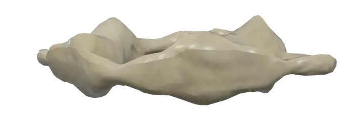
See it on the 3D body
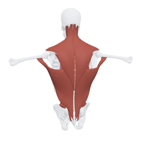
The atlas (C1 vertebra) lit on the 3D back, as in the back quiz. Turn the body, peel the layers, tap any structure for its name.
Quick answer
- Latin name
- Atlas
- Type
- Irregular bone (atypical cervical vertebra)
- Location
- Top of the neck, directly under the skull
The atlas is the first cervical vertebra (C1), a ring of bone at the top of the spine that carries the skull. Unlike other vertebrae, it has no body and no spinous process: two arches join two thick lateral masses, which hold the skull above and pivot on the axis (C2) below.
Where it is and what it touches
The atlas lies just below the base of the skull, deep at the top of the neck. Its name comes from the Titan of Greek myth who held up the sky: the atlas holds up the head.
It has two pairs of joints, plus a pivot in the middle:
- Above, its two concave superior articular facets cradle the occipital condyles of the skull at the atlanto-occipital joints. These joints mainly allow nodding, the "yes" movement, and a little side bending.
- Below, its flat inferior articular facets rest on the axis at the two lateral atlanto-axial joints.
- In front, the inner side of its anterior arch meets the dens of the axis, a peg of bone, at the median atlanto-axial joint.
The atlanto-axial joints are where the head turns from side to side, the "no" movement. Roughly half of the rotation of the neck happens here.
The bones of the spine page compares the atlas with the other 23 movable vertebrae.
Shape and landmarks
Seen from above, the atlas looks like a ring, wider from side to side than from front to back.
Anterior arch
- A short, curved bar at the front.
- The anterior tubercle on its front surface anchors the longus colli muscle and a ligament.
- The facet for the dens, on its back surface, is a small oval joint surface where the dens of the axis turns.
Posterior arch
- A longer, thinner bar at the back, curving around the spinal cord.
- The posterior tubercle in the midline replaces the spinous process found on other vertebrae.
- On its upper surface, behind each lateral mass, a groove for the vertebral artery marks where this artery curves over the arch on its way into the skull.
Lateral masses
- The thick parts at the sides of the ring, which carry the weight of the head.
- Each has a superior articular facet, oval and cupped, for the skull, and an inferior articular facet, round and nearly flat, for the axis.
- On the inner side of each mass, a small tubercle anchors the transverse ligament of the atlas, a strong band that crosses the ring behind the dens and holds it against the anterior arch.
Transverse processes and foramina
- The transverse processes of the atlas are wider than those of any other cervical vertebra; you can sometimes feel them deep between the angle of the jaw and the mastoid process behind the ear.
- Each one is pierced by a transverse foramen, through which the vertebral artery passes on its way up to the brain.
Vertebral foramen
The large central opening, divided by the transverse ligament: the dens sits in front, and the spinal cord and its coverings pass behind.
Why the atlas has no body
During development, the part that would have become the body of C1 joins the axis instead and becomes its dens. The atlas keeps only its arches and lateral masses, and the axis gains a peg that the atlas rotates around. This arrangement trades the stability of stacked bodies and discs for a wide range of rotation.
There is also no intervertebral disc between the skull and the atlas, or between the atlas and the axis. The first disc of the spine lies between C2 and C3.
Muscles that attach to the atlas
Several small and large neck muscles use the atlas as an anchor:
- The levator scapulae starts on the transverse processes of the upper cervical vertebrae, including the atlas, and runs down to the scapula; see the muscles of the shoulder.
- The splenius cervicis inserts on the transverse processes of the upper cervical vertebrae, including the atlas, and helps extend and turn the neck.
- The suboccipital muscles, deep under the base of the skull, also attach here: the rectus capitis posterior minor starts on the posterior tubercle, the obliquus capitis superior starts on the transverse process, and the obliquus capitis inferior inserts on it.
The large superficial muscles of the neck, such as the trapezius and the sternocleidomastoid, act on the head and neck from above and below but do not attach to the atlas itself.
In Anatomy Atlas 3D
In the app, the first vertebra is named "Atlas (C1 vertebra)". Tap it and turn the model to see the ring from above, then tap the axis below it to see the dens fit inside the anterior arch. Anatomy Atlas 3D is free on iPad and iPhone, works offline and shows English and Latin names.
Frequently asked questions
Why is C1 called the atlas?
After Atlas, the Titan of Greek myth who held up the sky. The C1 vertebra holds up the skull.
Does the atlas have a vertebral body?
No. Its body becomes the dens of the axis during development, so the atlas is a ring made of two arches and two lateral masses.
What movement happens between the skull and the atlas?
Mainly nodding, the "yes" movement. Turning the head side to side happens mostly at the joints between the atlas and the axis.
What holds the dens against the atlas?
The transverse ligament of the atlas, which crosses behind the dens and attaches to the inner sides of the lateral masses.
Is there a disc between C1 and C2?
No. The first intervertebral disc lies between C2 and C3.
Muscles that originate here
Muscles that insert here
From the anatomical data behind Anatomy Atlas 3D (Z-Anatomy, reviewed). In the app, tap the bone, then each muscle around it to open its card.
