
Muscle · Upper limb
Lumbricals of the hand: origin, insertion, action and innervation
The four lumbricals run from the flexor digitorum profundus tendons to the extensor expansions. They flex the knuckles. Nerves: median and ulnar.
Quick answer
- Latin name
- Musculi lumbricales manus
- Origin
- Tendons of the flexor digitorum profundus in the palm: first and second from the radial side of the tendons to the index and middle fingers; third and fourth from the facing sides of neighboring tendons
- Insertion
- Radial side of the extensor expansion (dorsal digital expansion) of fingers 2-5
- Action
- Flex the metacarpophalangeal joints and extend the interphalangeal joints of fingers 2-5
- Innervation
- First and second: median nerve (C8-T1). Third and fourth: deep branch of the ulnar nerve (C8-T1)
- Blood supply
- Branches of the superficial and deep palmar arches
See it on the 3D body
The lumbricals of the hand lit on the 3D forearm and hand, as in the forearm and hand quiz. Turn the body, peel the layers, tap any structure for its name.
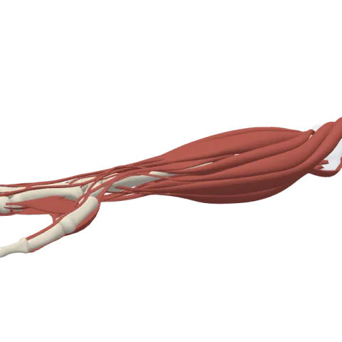
The lumbricals are four small, worm-shaped muscles in the palm that run from the tendons of the flexor digitorum profundus to the back of the fingers. They bend the knuckles while straightening the finger joints, and their nerve supply is split: the median nerve for the first and second, the deep branch of the ulnar nerve for the third and fourth.
Where it is
The lumbricals lie in the middle of the palm, under the palmar aponeurosis and the superficial palmar arch, right beside the four tendons of the flexor digitorum profundus. They are numbered from the index side: the first goes to the index finger, the fourth to the little finger. Their name comes from lumbricus, the Latin word for earthworm, which they resemble in shape and size.
Each lumbrical runs toward the web between two fingers, passes in front of the deep transverse metacarpal ligament and turns around the radial side (thumb side) of its finger to reach the back of the proximal phalanx. There it joins the extensor expansion, the flat sheet of tendon that the extensor digitorum spreads over the back of each finger.
They are unusual muscles: they have no attachment to bone. They start on one tendon and end on another, linking the flexor system of the palm to the extensor system on the back of the fingers.
Origin and insertion
- First lumbrical: from the radial side of the profundus tendon to the index finger.
- Second lumbrical: from the radial side of the profundus tendon to the middle finger.
- Third lumbrical: from the facing sides of the tendons to the middle and ring fingers.
- Fourth lumbrical: from the facing sides of the tendons to the ring and little fingers.
- Insertion: the radial side of the extensor expansion of fingers 2 to 5, over the proximal phalanx (see the phalanges of the hand).
The first two start from a single tendon each (unipennate); the last two start from two tendons each (bipennate).
What it does
The lumbricals pass in front of the metacarpophalangeal joints but join the extensor expansion behind the interphalangeal joints. As a result they do two things at once:
- Flex the metacarpophalangeal joints (the knuckles).
- Extend the proximal and distal interphalangeal joints (keep the fingers straight).
Together these give the "table-top" position: knuckles bent, fingers straight, as when a waiter balances a tray on flat fingers. Writing, dealing cards and many fine finger movements depend on this combination. The palmar interossei and the dorsal interossei help them with it.
When the ulnar nerve is damaged at the wrist, the third and fourth lumbricals lose their supply, and the ring and little fingers can drift into a clawed position, with the knuckles extended and the finger joints bent.
Innervation and blood supply
- First and second lumbricals: median nerve (C8-T1), through its digital branches in the palm.
- Third and fourth lumbricals: deep branch of the ulnar nerve (C8-T1).
The split roughly matches the profundus tendons they come from: the median side of the hand for the index and middle fingers, the ulnar side for the ring and little fingers. Their blood comes from branches of the superficial and deep palmar arches.
How to remember it
- Four earthworms on the tendons: picture four little worms lying on the profundus tendons in the palm.
- LOAF: the median nerve supplies the Lumbricals 1 and 2, Opponens pollicis, Abductor pollicis brevis and Flexor pollicis brevis. The rest of the small hand muscles belong to the ulnar nerve.
- The waiter's hand: knuckles bent, fingers straight under a tray. That is the lumbrical position.
- Always the thumb side: every lumbrical passes on the radial side of its finger.
The bones of the hand page shows the metacarpals they run between. Test yourself in the forearm and hand quiz.
Frequently asked questions
How many lumbricals are in the hand?
Four, one for each finger from the index to the little finger. The thumb has none.
What do the lumbricals do?
They flex the metacarpophalangeal joints and extend the interphalangeal joints of fingers 2 to 5, giving the table-top position of the hand.
What nerve supplies the lumbricals?
The first and second are supplied by the median nerve; the third and fourth by the deep branch of the ulnar nerve.
Where do the lumbricals originate?
On the tendons of the flexor digitorum profundus in the palm, not on bone.
Where do the lumbricals insert?
On the radial side of the extensor expansion on the back of each finger.
