
Muscle · Upper limb
Flexor digitorum superficialis: origin, insertion, action and innervation
The flexor digitorum superficialis runs from the elbow to the middle phalanges of fingers 2-5. It bends their middle joints. Nerve: median nerve.
Quick answer
- Latin name
- Musculus flexor digitorum superficialis
- Origin
- Humero-ulnar head: medial epicondyle of the humerus (common flexor origin) and coronoid process of the ulna. Radial head: upper half of the anterior border of the radius
- Insertion
- Sides of the middle phalanges of fingers 2-5 (phalanges of the hand)
- Action
- Flexes the proximal interphalangeal joints of fingers 2-5; also flexes the metacarpophalangeal joints and helps flex the wrist
- Innervation
- Median nerve (C7-T1)
- Blood supply
- Branches of the ulnar and radial arteries
See it on the 3D body
The flexor digitorum superficialis lit on the 3D forearm and hand, as in the forearm and hand quiz. Turn the body, peel the layers, tap any structure for its name.
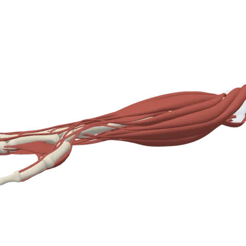
The flexor digitorum superficialis (FDS) is the broad muscle in the middle layer of the front of the forearm whose four tendons bend the index, middle, ring and little fingers at their middle joints. It starts near the elbow from the humerus, the ulna and the radius, and it is supplied by the median nerve.
Where it is
The FDS forms the intermediate layer of the anterior forearm on its own. The four superficial muscles, the pronator teres, the flexor carpi radialis, the palmaris longus and the flexor carpi ulnaris, lie in front of it. The deep layer, with the flexor digitorum profundus and the flexor pollicis longus, lies behind it. Despite its name, it is mostly covered by other muscles near the elbow and only comes close to the skin in the lower forearm.
Its two heads are joined by a fibrous arch. The median nerve and the ulnar artery pass under this arch, and the median nerve then runs down the forearm stuck to the deep surface of the muscle until it appears at its outer edge just above the wrist.
Above the wrist, the FDS gives off four tendons that pass through the carpal tunnel in two rows: the tendons for the middle and ring fingers in front, those for the index and little fingers behind. In each finger, at the level of the proximal phalanx, the tendon splits into two slips. The profundus tendon passes through the gap, and the two slips go around it to attach on either side of the middle phalanx.
Origin and insertion
- Humero-ulnar head: the medial epicondyle of the humerus through the common flexor tendon, the ulnar collateral ligament of the elbow and the coronoid process of the ulna.
- Radial head: a thin sheet from the upper half of the anterior border of the radius.
- Insertion: the two sides of the shaft of the middle phalanges of fingers 2 to 5 (see the phalanges of the hand).
What it does
- Flexes the middle finger joints: its main action is flexion of the proximal interphalangeal (PIP) joints of the four fingers.
- Flexes the knuckles: it continues to pull on the metacarpophalangeal joints as the fingers close.
- Helps flex the wrist: its tendons cross the front of the wrist, so it assists the wrist flexors.
Its slips for each finger are more independent than those of the profundus, which lets you bend one finger at a time. In a strong grip it works alongside the profundus.
To test it, hold the other fingers fully straight. Because the profundus bellies for those fingers are joined, this stops the profundus from working, and the finger being tested bends at the middle joint while its tip stays loose. In some people the slip for the little finger is small or missing, so that finger may not bend well in this test.
Innervation and blood supply
The FDS is supplied by the median nerve (C7-T1). The nerve gives its branches to the muscle near the elbow, then travels on its deep surface down the forearm. Its blood comes from branches of the ulnar artery and the radial artery.
How to remember it
- Superficialis stops in the middle: its tendons end on the middle phalanges, so it bends the middle joints.
- Single and pair: the Superficialis has a Single nerve (median); the Profundus has a Pair (median and ulnar).
- Middle pair up front: in the carpal tunnel, the tendons of the two middle fingers (middle and ring) lie in front of the outer two (index and little).
- Three bones, two heads: one head starts on the humerus and the ulna, the other on the radius.
Compare it with the other layers on muscles of the forearm, then test yourself in the forearm and hand quiz.
Frequently asked questions
What does the flexor digitorum superficialis do?
It flexes the proximal interphalangeal joints of fingers 2 to 5, the middle joints of the fingers. It also helps flex the knuckles and the wrist.
Where does the flexor digitorum superficialis insert?
On the two sides of the middle phalanges of the index, middle, ring and little fingers. Each tendon splits so the profundus tendon can pass through.
What nerve supplies the flexor digitorum superficialis?
The median nerve, from roots C7 to T1.
How do you test the flexor digitorum superficialis?
Hold the other fingers straight to block the profundus, then ask the person to bend the finger being tested. It should bend at the middle joint with a loose fingertip.
What passes under the arch of the flexor digitorum superficialis?
The median nerve and the ulnar artery, on their way from the elbow into the forearm.
